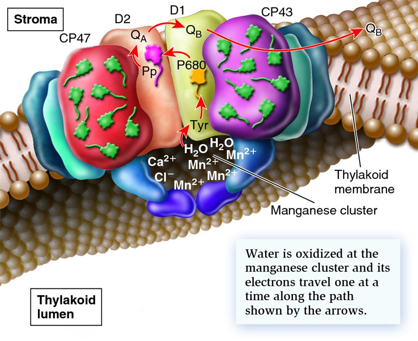
Schematic drawing showing the path of electron flow from water to Q B. The CP47 and CP43 protein subunits wrap around D1 and D2 so that pigments in CP47 and CP43 can transfer energy to P680 by resonance energy transfer.
The oxidation of water occurs in a region called the manganese cluster. This site is located on the side of D1 that faces the thylakoid lumen. The manganese cluster has four Mn2+,, one Ca2+, and one Cl?. Two water molecules bind to this site. D1 catalyzes the removal of four electrons from the two water molecules to create four H+ and O2. The electrons are transferred, one at a time, to a tyrosine (Tyr) in D1 and then to an oxidized pigment molecule (P680+) to produce P680. When the electron on P680 becomes excited, usually by resonance energy transfer, it then moves to the primary electron acceptor, which is an organic molecule called pheophytin (Pp) that is permanently bound to photosystem II. Pheophytin transfers its electron to a plastoquinone molecule, designated Q A, which is also permanently bound to photosystem II. Next, the electron is transferred to another plastoquinone molecule designated Q B, which can accept two high-energy electrons and bind two H+. Q B can diffuse away from the reaction centre.
In 2004, So Iwata, James Barber, and colleagues determined the three-dimensional structure of photosystem II by using a technique called X-ray crystallography. In this method, researchers must purify a protein or protein complex and expose it to conditions that cause the proteins to associate with each other in an ordered array. In other words, the proteins form a crystal. When a crystal is exposed to X-rays, the resulting diffraction pattern can be analyzed mathematically to determine the three-dimensional structure of the crystal's components. Major advances in this technique over the last couple of decades have enabled researchers to determine the structures of relatively large macromolecular machines, such as photosystem II and ribosomes.
Photosystems II and I Work Together to Produce ATP and NADPH Let's now consider what happens to the high-energy electrons that are transferred to the primary electron acceptor in photosystem II (Figure 8.9). After electrons reach Q B, they enter an electron transport chain located in the thylakoid membrane. The electron transport chain functions similarly to the one found in mitochondria. From Q B, electrons go to a cytochrome complex, then to plastocyanin (Pc), a small protein, and then to a pigment molecule in the reaction centre of photosystem I. Along the journey from photosystem II to photosystem I, the electron releases some of its energy at particular steps and is transferred to the next component that has a higher electronegativity. The energy that is released is harnessed to move H+ into the thylakoid lumen. One result of the electron movement is to establish an H+ electrochemical gradient. Additionally, the splitting of water also adds H+ into the thylakoid lumen. The synthesis of ATP in chloroplasts is achieved by a chemiosmotic mechanism similar to that used to make ATP in mitochondria.
Related Images
219
836
1607
181
635
195
88
1491
271
138196
Add Comment
0 Comment
Explore
Post your homework questions and get free online help from our incredible volunteers