× Didn't find what you were looking for? Ask a question
Top Posters
Since Sunday
g
3
3
2
J
2
p
2
m
2
h
2
s
2
r
2
d
2
l
2
a
2
New Topic  
mcats1675 mcats1675
wrote...
12 years ago
im doing a project for class about the process of urine formation in the nephron.  i understand how it is all done, but im having problems translating it from what i understand to paper.  what is the most simple way to do so?  lamens terms please!
Read 4920 times
4 Replies

Related Topics

Replies
wrote...
Staff Member
12 years ago
Hey,

Urine is produced in the kidneys. Once it is produced, it is carried to the urinary bladder by the ureters. But before the kidneys can actually produce urine and release it into the urinary bladder, blood leading to the kidneys is filtered. Arteries that lead to the kidney are called afferent arterioles.  The are called arterioles because this word represents "baby arteries". The blood pressure here is relatively high. Remember, blood contains cells, water, proteins, ions, sugars, amino acids, etc. You don't want to lose all those good stuff, but rather you want to get rid of the toxins found in the blood and excess water (perhaps from drinking too much water). Once in the kindey, the blood goes through this mesh filter called the glomerulus. Since the kidneys is made up of millions of nephrons, each nephron has its own glomerulus. Below is an image of a single glomerulus:

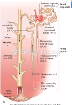
http://www.siumed.edu/~dking2/crr/images/corp5.jpg

Below is an image of a nephron; remember, these things are tiny and there are millions of them:

http://www.cic-caracas.org/departments/science/images/08nephron1.jpg

Anyways, the nephron has different sections. According to the image above, the first section is the proximal tubules. Because the glomerulus isn't perfect, if may not do a good job filtering everything at the Bowman's capsule and it may allow things like sugars and proteins to pass through. So, in the nephron, the primary function of the proximal tubule is to reabsorb ions, organic molecules, and water. This process is called reabsorption and there are four main steps in urine formation, with reabsorption being one of them. What ever is left after this original section goes into the loop of Henle (that long part that looks like a large loop). In other words, it's the hairpin-shaped segment of the nephron. Here, more water and ions (such as sodium and chloride) is reabsorbed into blood vessels that surround the loop. This can be done through either active transport or passive transport - depending on what's being reabsorbed.

Next, urine moves through the distal tubule, the last segment of the nephron, and finally into the collecting ducts. As the name suggests, the collecting ducts collect urine from the million nephrons that, in turn, merge in the pelvis of the kidney and flow into the ureters, which in turn lead to the bladder.
- Master of Science in Biology
- Bachelor of Science
marie Author
wrote...
12 years ago
thanks for the help....just one more question....what nutrients and products are removed in each step of the production process in the nephron?
wrote...
Educator
12 years ago
Sure, check it out:

 Attached file 
Thumbnail(s):
You must login or register to gain access to this attachment.
lagrangeboo Author
wrote...
12 years ago
The organs, tubes, muscles, and nerves that work together to create, store, and carry urine are the urinary system. The urinary system includes two kidneys, two ureters, the bladder, two sphincter muscles, and the urethra.

How does the urinary system work?


Front view of urinary tract.

Your body takes nutrients from food and uses them to maintain all bodily functions including energy and self-repair. After your body has taken what it needs from the food, waste products are left behind in the blood and in the bowel. The urinary system works with the lungs, skin, and intestines—all of which also excrete wastes—to keep the chemicals and water in your body balanced. Adults eliminate about a quart and a half of urine each day. The amount depends on many factors, especially the amounts of fluid and food a person consumes and how much fluid is lost through sweat and breathing. Certain types of medications can also affect the amount of urine eliminated.

The urinary system removes a type of waste called urea from your blood. Urea is produced when foods containing protein, such as meat, poultry, and certain vegetables, are broken down in the body. Urea is carried in the bloodstream to the kidneys.

The kidneys are bean-shaped organs about the size of your fists. They are near the middle of the back, just below the rib cage. The kidneys remove urea from the blood through tiny filtering units called nephrons. Each nephron consists of a ball formed of small blood capillaries, called a glomerulus, and a small tube called a renal tubule. Urea, together with water and other waste substances, forms the urine as it passes through the nephrons and down the renal tubules of the kidney.

From the kidneys, urine travels down two thin tubes called ureters to the bladder. The ureters are about 8 to 10 inches long. Muscles in the ureter walls constantly tighten and relax to force urine downward away from the kidneys. If urine is allowed to stand still, or back up, a kidney infection can develop. Small amounts of urine are emptied into the bladder from the ureters about every 10 to 15 seconds.

The bladder is a hollow muscular organ shaped like a balloon. It sits in your pelvis and is held in place by ligaments attached to other organs and the pelvic bones. The bladder stores urine until you are ready to go to the bathroom to empty it. It swells into a round shape when it is full and gets smaller when empty. If the urinary system is healthy, the bladder can hold up to 16 ounces (2 cups) of urine comfortably for 2 to 5 hours.

Circular muscles called sphincters help keep urine from leaking. The sphincter muscles close tightly like a rubber band around the opening of the bladder into the urethra, the tube that allows urine to pass outside the body.

Nerves in the bladder tell you when it is time to urinate, or empty your bladder. As the bladder first fills with urine, you may notice a feeling that you need to urinate. The sensation to urinate becomes stronger as the bladder continues to fill and reaches its limit. At that point, nerves from the bladder send a message to the brain that the bladder is full, and your urge to empty your bladder intensifies.

When you urinate, the brain signals the bladder muscles to tighten, squeezing urine out of the bladder. At the same time, the brain signals the sphincter muscles to relax. As these muscles relax, urine exits the bladder through the urethra. When all the signals occur in the correct order, normal urination occurs.


New Topic      
Explore
Post your homework questions and get free online help from our incredible volunteers
  1112 People Browsing
 121 Signed Up Today
Related Images
  
 75
  
 480
  
 188
Your Opinion
Who will win the 2024 president election?
Votes: 3
Closes: November 4