|
Hey Karim89,
All special sense organs develop mostly from ectoderm. The ear begins its development together with the eye early during the fourth week of embryogeneis. The development of the ear involves ectoderm (surface ectoderm and neuroectoderm), mesoderm, and endoderm. The sensory components of the ear develop by the 20th week of embryogensis and the accessory structures by week 32. The ear of an adult structurally and functionally consists of an external ear, a middle ear, and an inner ear. Each of these has a separate embryonic origin.
Inner Ear
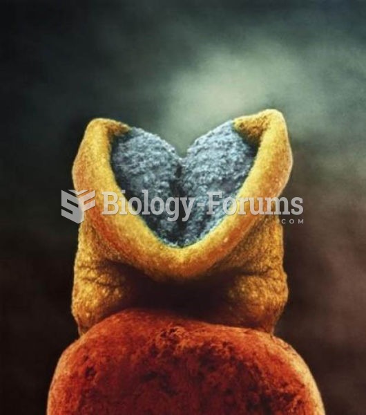
The inner ear begins to develop early in the third week when a plate of surface ectoderm called the otic placode appears on the lateral side of the rhombencephalon. The otic placode soon invaginates to form an otic pit. Toward the end of the fourth week, the outer edges of the invaginated otic pit converge and fuse to form an otocyst (otic vesicle). The otocyst soon separates from the surface ectoderm and then differentiates to form a dorsal utricular portion and a ventral saccular portion.
Three diverticula extend outward from the utricular portion and develop into the semicircular canals that later function in balance and equilibrium. A tubular diverticulum, called the cochlear duct, extends in a spiral fashion from the saccular portion and becomes the membranous portion of the cochlea. The organ of Corti, the functional portion of the cochlea, differentiates from cells along the wall of the cochlear duct. During the sixth week, the neuroectoderm of the developing brain differentiates into the spinal and vestibular ganglia, and their sensory nerves that innervate the inner ear extend toward the developing air cells.
The mesodermal tissue (mesenchyme) that surrounds the differentiating otocyst soon forms a cartilaginous otic capsule. As the otocyst and surrounding otic capsule grow, vacuoles containing the fluid perilymph develop within the otic capsule. The vacuoles soon enlarge and unite to form the perilymphatic space that divides into the scala tympani and the scala vestibuli. The cartilaginous otic capsule eventually ossifies to form the bony (osseous) labyrinth of the inner ear.
Thus, the sacculus, utriculus, cochlea, semicircular canals, and the auditory nerve and ganglion develop from the otic placode and vesicle (surface ectoderm). Secondary supporting investments of the otic vesicle come from mesoderm.
Middle Ear
The lining of the middle ear chamber, or tympanic cavity, develops from foregut endoderm associated with the first pharyngeal pouch. The auditory ossicles that amplify incoming sound waves develop from the cartilages of the first and second pharyngeal arch. The tympanic cavity enlarges, surrounds, and encloses the developing ossicles. The connection of the tympanic cavity to the pharynx gradually elongates into the auditory (eustachian) tube. The auditory tube remains patent throughout life and is important in maintaining equilibrium of air pressure between the pharyngeal and tympanic cavities.
External Ear
The external ear includes the fleshy auricle (pinna) attached to the side of the head and the tubular external auditory meatus that continues into the temporal bone of the skull. The external auditory meatus develops from the surface ectoderm that covers the dorsal end of the first pharyngeal grove. A solid epithelial plate meatal plug soon develops at the bottom of the funnel-shaped pharyngeal groove. The meatal plug contributes to formation of the inner wall of the external auditory meatus and to the tympanic membrane (eardrum). The inner surface of the eardrum (tympanum) develops from foregut endoderm associated with the first pharyngeal pouch. The outer surface of the eardrum develops from surface ectoderm. Mesoderm separates the two layers of this first branchial membrane.
The auricle develops from six separate swellings of surface ectoderm called auricular hillocks that develop around the first branchial groove. The auricular hillocks enlarge, unite, and become supported by mesodermal mesenchyme that differentiates into fibrous tissue. The auricles migrate superiorly as the face of the embryo and fetus develops. The auricles have ascended to the dorsoventral level of the eyes by thirty-two weeks.
-------------------
In terms of the video/animation, i remember having videos on these a while back, I will check and get back to you ASAP
|

 Quick Reply