|
|
A free membership is required to access uploaded content. Login or Register.
Transportin-Humans
|
|
Uploaded: 7 years ago
Contributor: Guest
Category: Biology
Type: Lecture Notes
Tags: valves, arteries, artery, sino-atrial, elastic, tricuspid, coronary, cardiac, ventricle, stroke, tissue, circulatory, pacemaker, ventricles, double
Rating:
N/A
|
Filename: Transportin-Humans.docx
(632.6 kB)
Page Count: 1
Credit Cost: 2
Views: 179
Last Download: N/A
|
Transcript
The Heart
Cardiac muscle that contracts and relaxes rhythmically.
Has its own blood supply for glucose and oxygen.
Vessels are called coronary arteries and veins.
Blockage of these vessels with cholesterol results in coronary heart disease which leads to myocardial infarction (heart attack).
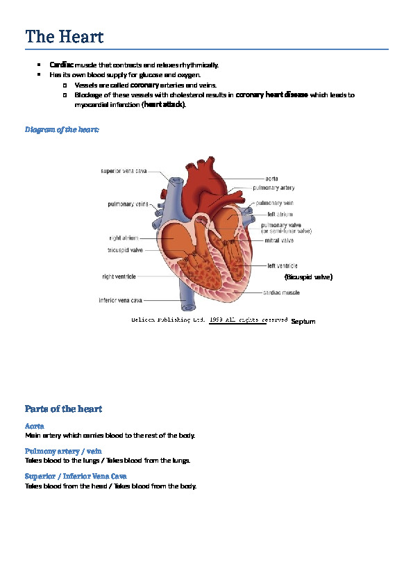
Diagram of the heart:
center339090001173480413004000
1285240425323000
4743450135890(Bicuspid valve)
00(Bicuspid valve)
487997587630Septum
00Septum
38906455334000
Parts of the heart
Aorta
Main artery which carries blood to the rest of the body.
Pulmony artery / vein
Takes blood to the lungs / Takes blood from the lungs.
Superior / Inferior Vena Cava
Takes blood from the head / Takes blood from the body.
Heart beat
Called a cardiac cycle
Volume in one cycle = stroke volume
Volume in one minute = stroke volume x beats per minute = cardiac output
General diastole
Both atria relax and blood flows into all four chambers.
Bicuspid and tricuspid valves open.
Semi-lunar valves are closed.
Artrial systole
Both atria contract, squeezing blood into the ventricles and raising the pressure in the ventricles.
Bicuspid and tricuspid valves still open.
Semi-lunar valves still closed
Ventricular systole
Ventricles contract, squeezing blood into the arteries to lungs and the rest of the body.
Bicuspid and tricuspid valves close.
Semi-lunar valves open.
1355725-381000
-190506286500
Heart valves
Tendons support the valves and ensure that they do not collapse in the wrong direction.
Tricuspid valve (Atrioventricular valve)
Between RIGHT atrium and RIGHT ventricle.
Bicuspid valve (Atrioventricular valve)
Between LEFT atrium and LEFT ventricle.
Semi-lunar valves
Between RIGHT ventricle and PULMONARY artery
Between LEFT ventricle and AORTA.
Control of the heartbeat
Specialized muscle cells can conduct electrical impulses and give the heart the signal to start beating.
Most important patch of these cells is the sino-atrial node (pacemaker) which is found in the right atrium.
Other patches:
Atrioventricular node which is lower down in the right atrium.
Pukyne fibres in the bundle of His, which is near the bottom of the septum.
Signals of the heartbeat:
First electrical impulse from the sino-atrial node, after which both atria contract.
Signal then moves to the atrioventricular node, which delays the conduction for the atria to relax.
Signal then moves to the Purkyne fibres, after which both ventricles contract.
Role of the brain:
Sends nerve impulses to the sino-atrial node to speed up or slow down heart rate according to the body’s metabolic needs (According to CO2 and O2 concentrations as well as energy being used).
If the sino-atrial node stops working, an artificial pacemaker may be inserted into the heart to keep the heart beating regularly.
Diseases of heart and arteries (Cardiovascular diseases)
Atherosclerosis
Caused by the hardening of the arteries which is usually due to the build up of cholesterol in the lining.
Very common in the coronary arteries around the heart, causing them to narrow
This is called Coronary Heart Disease or CHD.
Coronary Heart Disease (CHD)
Can lead to the blockage of the blood flow to a section of the heart.
When this occurs the heart cannot get glucose or oxygen and a myocardial infarction (heart attack) occurs.
This may be fatal.
Risk Factors:
Genetic predisposition
High-cholesterol diet
Obesity
Stress
Smoking tobacco
Little/no exercise
Blood vessels
514350-190500133350362331000
2733675-296545Diagram of cross-section of artery or vein:
(Thickness of layers differ)
00Diagram of cross-section of artery or vein:
(Thickness of layers differ)
Arteries
251396538735Connective tissue made up mainly of elastic fibres
Smooth muscle
and elastic fibres
Epithelium
One cell thick lining
00Connective tissue made up mainly of elastic fibres
Smooth muscle
and elastic fibres
Epithelium
One cell thick lining
Away from the heart/organ etc.
No valves
Elastic for smooth blood flow
Strong walls
High pressure
Pulses
Veins
To the heart/organs etc.
Semi-lunar valves
Blood is pushed steadily through by the contraction of surrounding muscles
If no movement, blood clots, which can then shoot up into the brain and death is imminent.
Low pressure
Wider lumen
168275063500
3009265100965Endothelial cell
00Endothelial cell
259334023876000Capilaries
One cell thick
Very thin walls
Penetrate entire body
Carry materials to and from cells in the bod
Efficient exchange between blood and tissues
Circulation
Humans have a double circulatory system
This means that blood flows through the heart twice per one cycle through the body.
The double circulatory system consists of:
Pulmonary circuit – from the heart to the lungs and then back to the heart.
Systemic circuit – from the heart to the rest of the body and back to the heart.
39814509144000This is represented on the diagram alongside:
Blue – Deoxygenated blood
-171450166497000Red – Oxygenated blood
-438150995045Superior
00Superior
-4381502443480Inferior
00Inferior
Important arteries and veins:
|
|
Comments (0)
|
Post your homework questions and get free online help from our incredible volunteers
|