|
CAN ANYONE HELP ME WITH E. PLEASE?!?
Situation: Melissa is practicing her golf swing at a driving range.
(Part One): Muscles, Joints and Bones Anatomy [50 total points possible]
a. Which bones of the body are used to go from the anatomical position to holding the golf club in front of her with both hands as seen in the picture above?
The bones that are involved in the movement of the upper body from the anatomical position to the position of holding a golf club with both hands at the front of the body involve all sixty four (64) bones composed from two sections on each side of the upper body:
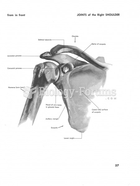
Pectoral Girdle, “Shoulder bones.” The Pectoral Girdle is made up of two bones on each individual side of the upper body which include both the Scapula bone & clavicle bone. The total amount of bones in the Pectoral Girdle is four (4).
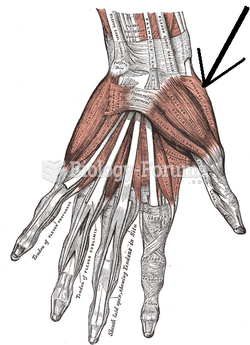
The bones that compose the arms, wrists, hands and fingers are what make up the upper limb. The upper limb consists of the Humerus, Radius, Ulna, Carpals (8), Metacarpals (5), and Phalanges (14) bones. Total number of bones of both sides of the upper limbs is sixty (60).
For the body to move from the anatomical position to holding the golf club in the front of the body using both hands all of the bones of each upper limb would be moved simultaneously from muscular force. The Humerus, Radius and Ulna would be moved in a forward position and then to a position that is forward and elevated.
From the moment the club is lifted into the air, the Humerus is lifted upward by muscular force and free movement of the ball and socket of the shoulder. While the club is lifted, the angle of the drive is created by the posture of the golfer. The Radius and Ulna are moved from the anatomical position to an upward position and to a different angle towards the golfer’s upper left or right side of the body. The Carpals, Metacarpals, and Phalanges help create the angle of the golfer’s drive. These bones will establish the angle of the drive by the position that they are in while in the upward position until the moment that the club makes contact with the ball. The Phalanges and Metacarpals are also responsible for providing the required grip of the club.
Phalanges (fingers)
Metacarpals (hand)
Carpals (wrist)
Radial and Ulna (forearm)
Olecranon and Trochlear (elbow)
Humerus (upper arm)
Acromion, Clavicle and Scapula (shoulder)
b. Which joints are used, and what is the type of action for each joint (i.e. flexion, extension, etc.)?
Involved in the act of moving the upper body from the anatomical position to the position of holding the golf club in front of the body with both hands requires the usage of multiple joints working simultaneously. From the moment the upper body moves from the anatomical position to the time the golf club is being held in the front of the body roughly four individual joints have been utilized, each of which have performed different kinds of movements.
The joints that are used are known as synovial joints. These are different than other joints in that they allow the joint to be freely movable by means of a synovial joint cavity that exists between the connected bones. This freedom of movement allows for the bones to move at different angles; which include gliding, angular, rotation and special movements. Of these four styles of movements, each has a variety of different functions that they offer for different movements for various bone structures and make it possible for us to move in the ways that we do.
The synovial joints each have different structures and functions dependant on the location of the joint and bone. When the golfer is at the anatomical position the joints are at the normal, resting position. At this time there is no exertion being placed on the joints. When the golfer swings the golf club all sixty of the combined upper limb bones & Pectoral Girdle are moved to different positions by means of the synovial joints. The Humerus bones are moved from the anatomical position of rest to a drawn back position by gliding of the scapula and clavicle and by both extension and flexion from joints.
If the golfer is right handed for instance and the high point of the swing results in the club being brought toward the upper right side of the body then the left arm (Humerus, Ulna, and Radius) is allowed to go to that position by the hinge joints and pivot joints of the elbow by movement known as extension. The golfer’s right arm (Humerus, Ulna, and Radius) goes through flexion because that arm is being pulled upward while being bent upwards at the elbow’s hinge and pivot joints and from the ball & socket joints at the Humerus and Scapula. There is also a slight rotational movement in the upper limb ball & socket joints that help let the Humerus and Scapula allow the upper limbs to move in that manner. The positions of the hands are created by the carpometacarpal joints allowing the hands to be at either the positions of pronation or supination.
The movement downwards toward the golf ball involves the same joints but the movement changes. The left arm goes through flexion and the right arm goes through extension. At the moment that the golf club is being held in front of the body both of the upper limbs (Humerus) continue to endure flexion. The synovial joints in the hands and wrists (Condlyloid joint, and Saddle joints) throughout the entire movement have gone through abduction (movement away) and adduction (movement toward) as well as flexion & extension at the wrists. The carpometacarpal joints in the hands allow for the oppositional movement of the hands and Phalanges by allowing the golfer to grip the golf club.
Wrist Joint – Condyloid and is biaxial (around two axis, perpendicular to each other). Flexion, extension, abduction, adduction.
Elbow – Hinge and is uniaxial (around one axis, in one place).
Flexion and extension.
Shoulder –Ball and joint and is multiaxial (around many axes).
Flexion, extension, abduction, adduction, rotation, circumduction.
Bones; phalanges, radials, ulna, Humerus
c. Which muscles are required to complete the stance?
Muscles that move the hand and wrist:
Flexor carpi radialis – flexes the hand and forearm.
Palmaris longus – Flexes the hand.
Flexor carpi radialis longus – Extends the wrists and hand and abducts the hand.
Extensor carpi radialis brevis – Extends the hand.
Extensor carpi ulnaris – Extends and adducts the wrist and hand.
Flexor digitorum profundus – Flexes the distal joints of the fingers.
Flexor digitorum superficialis – Flexes the fingers.
Extensor digitorum- Extends the fingers.
Muscles that move the forearm:
Biceps brachi – Flexes the forearm at elbow, rotates the hand laterally.
Brachialis – Flexes the forearm at the elbow.
Brachioradialis – Flexes the forearm at the elbow.
Triceps brachil – Extends the lower arm at the elbow.
Pronator teres – Pronates the forearm.
Pronator quadratus – Pronates the forearm.
Supinator – Supinates the forearm.
Muscles that move the upper arm:
Pectoralis major – Flexes and adducts the upper arm and draws it anteriorly across the chest.
Lattissimus dorsi - Extends and adducts the upper arm posteriorly.
Deltoid – Assists in flexing, extending and adducting the upper arm.
Coracobrachialis - Assists in flexing and rotating the arm medially and in adduction.
Supraspinatrust (rotor cuff) – Assists in abducting the arm.
Teres minor (rotor cuff) – Rotates the arm outward.
Teres major – Assists in extending, adducting and rotating the arm medially.
Infraspinatrust (rotator cuff) – Rotates the arm laterally and outwardly.
Subscapularist (rotator cuff) – Rotates arm medially.
(Part 2): Nervous System and Muscle Physiology [40 total points possible]
d. What occurs from the moment Melissa decides to swing to actually completing the act? Detail the steps needed for the thought to go from the first neuron to the second, and then from the last neuron to one of the muscles you identified in Part 1c above.
It starts with sensory function that begins from being detected by the stimuli being produced by traveling to the cranial nerves. Then we go into the integrative function going to the brain to analyze what it wants to do. After this, we go into the last phase known as the motor function by traveling back through the cranial and spinal nerves which is going to incorporate the radial nerves then it passes down the posterial lateral surface of the arm and forearm supplying all of the external and extensor muscles of the arm, forearms, and hands, Once it has reached all these phases the action of swinging takes place.
e. Which nerve directly innervates the muscle you chose in Part 2d above? [3 points possible]
f. Explain the Sliding Filament Theory. [12 points possible]
Sliding filament Theory is based on the observation that the muscle fibers are composed of two types of filaments: thick filaments and thin filaments. Muscle fibers are made up of repeating units of sarcomere. Each sarcomere contains overlapping thick and thin filaments. In the middle of the sarcomere are thick filaments, which makes up the A band. The length of the A band does not change during contraction. The thin filaments are interlaced between the thick filaments and also overlap with the lateral portion of the thick filaments. The area of overlap is called the zone overlap. The myosin head of the thin filament can bind to the active site of the thick filament. The contraction of the muscle requires, then, movement of the thin filaments toward the thick filament medially. This is where the calcium ions are required. Calcium ions can bind to troponin, a molecule on the thick filament near the actice sites where myosin head of the thin filament can potentially bind. Due to binding of calcium, troponin changes shape and moves the active site so that it can interact with myosin head of the thin filament. Further, ATP is required for the power stroke of the myosin head, which can then move toward the center of the sarcomere. When another ATP binds to the myosin head, the active site and myosin head of the thin filament detach. Because of this, the active site is able to form another cross bridge, allowing further muscle contraction. Hence, during the muscle contraction of the zone of overlap lengthens while the H zone, the part of sarcomere composed of thick filament only shortens.
|
 Quick Reply