|
Replies
|
wrote...
|
|
|
10 years ago
|
Like most excitable cells, muscle fibers respond to the excitation signal with a rapid depolarization which is coupled with its physiological response: contraction.
Cellular Resting Potential
If we remember that myofibers are basically water with some dissolved ions separated from the extracellular space, which is also mostly water with some dissolved ions, then the presence of a resting potential may make more sense. In much the same way as a battery creates an electrical potential difference by having different concentrations of ions at its two poles, so does a muscle cell generate a potential difference across its cell membrane. The ATP driven sodium-potassium pump maintains an artificially low concentration of sodium and high concentration of potassium in the intracellular space, which generates a resting potential difference on the order of -75 mV.
Depolarization
Depolarization is achieved by other transmembrane channel proteins. When the potential difference near these voltage sensitive proteins reaches a threshold level, the protein undergoes a magical conformational change that makes the membrane permeable to sodium. Extracellular sodium immediately rushes in, drawn by both the charge difference and concentration gradient, and locally depolarizes the cell. Almost immediately, potassium also moves along in concentration gradient - out of the cell -- and the membrane potential is restored.
As an interesting side note, this is the mechanism by which potassium chloride is used to induce cardiac arrest: by eliminating the potassium concentration gradient, the depolarized cardiac muscle cells are unable to repolarize for their next beat.
Coordination of Depolarization
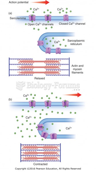
This depolarization is an extremely localized phenomenon, depending on diffusion over a few milliseconds. Some system is required to carry this signal to the myofibrils deep within the cell body. The sarcolemma, or cell membrane, invaginates to form a network of transverse (or T-) tubules that span the cross section of each fiber, transmitting the depolarization signal uniformly throughout the cell.
From Depolarization to Contraction
Contraction is regulated by calcium ion concentration. In the resting state, a fiber keeps most of its intracellular calcium carefully sequestered in an extensive system of vessicles known as the sarcoplasmic reticuluum. There are at least two receptors in the chain between depolarization and calcium release. Once released, calcium binds to troponin, opening the myosin binding sites on filamentous actin, and force is produced.
|
|
|
|
|
|
Mastering in Nutritional Biology Tralalalala 
|
|
|
wrote...
|
|
|
10 years ago
|
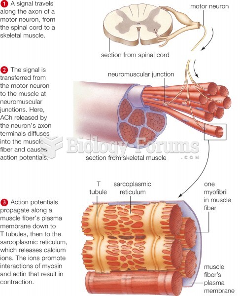
This process is fundamental to muscle physiology, whereby the electrical stimulus is usually an action potential and the mechanical response is contraction.
In skeletal muscle, E-C coupling relies on a direct coupling between key proteins, the sarcoplasmic reticulum (SR) calcium release channel (identified as the ryanodine receptor, RyR) and voltage-gated L-type calcium channels (identified as dihydropyridine receptors, DHPRs). DHPRs are located on the sarcolemma (which includes the surface sarcolemma and the transverse tubules), while the RyRs reside across the SR membrane. The close apposition of a transverse tubule and two SR regions containing RyRs is described as a triad and is predominantly where E-C coupling takes place. E-C coupling proceeds as follows:
1) The membrane potential of a skeletal muscle cell is depolarised by an action potential (e.g. from synaptic activation from an alpha motor neuron)
2) This depolarisation activates voltage-gated DHPRs
3) This activates RyR type 1 via physical linkage (involving conformational changes that allosterically activates the RyRs)
4) As the RyRs open, calcium is released from the SR into the local junctional space, which then diffuses into the bulk cytoplasm to cause a calcium transient. Note that the SR has a large calcium buffering capacity partially due to a calcium-binding protein called calsequestrin
5) The calcium released into the cytosol binds to Troponin C by the actin filaments, to allow cross-bridge cycling, producing force and, in some situations, motion
6) The sarco/endoplasmic reticulum calcium-ATPase (SERCA) actively pumps calcium back into the SR
7) As calcium declines back to resting levels, the force declines and relaxation occurs
|
|
|
|
|
|
|
 Quick Reply