Transcript
Cytoskeleton: a network of proteinaceous fibers that organizes structures and activities in the cell, anchor organelles, and help to support the cell and maintain its shape. It is more dynamic than an animal skeleton, however, it can be quickly dismantled in one part of the cell and reassembled in a new location, changing the shape of the cell. Some cytoskeleton structures interact with motor proteins in eukaryotic flagella and cilia to produce cell motility. It is composed of microtubules, microfilaments, and intermediate filaments.
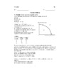
Microfilaments:
Are solid rods
is a twisted double chain of actin subunits
Besides occurring as linear filaments, it can form structural networks when certain proteins bind along the side of such a filament and allow a new filament to extend as a branch.
Are the thinnest type of fiber
In contrast to the compression-resisting role of microtubules,
the structural role of microfilaments in the cytoskeleton is to bear tension (pulling forces).
Microtubules: Are hollow rods made of ?- and ?- tubulin. They shape and support the cell and also serve as tracks along which organelles equipped with motor proteins can move.
-guide vesicles from the ER to the Golgi apparatus and from the Golgi to the plasma membrane. -Microtubules are also involved in the separation of chromosomes during cell division
intermediate filaments
-play an important role in reinforcing the shape of a cell and fixing the position of certain organelles.
-For instance, the nucleus typically sits within a cage made of intermediate filaments, fixed in location by branches of the filaments that extend into the cytoplasm.
-are more permanent fixtures of cells than microfilaments and microtubules, which are often disassembled and reassembled in various parts of a cell.
Tubulin: All eukaryotic cells have microtubules, hollow rods constructed from globular proteins called tubulins.
Actin: make up the microfilament (also called actin filaments). Microfilaments are solid rods about 7 nm in diameter. They are built as a twisted double chain of actin subunits
motor protein: are responsible for the undulations of cilia and flagella. Actin and myosin proteins are responsible for the contraction of muscles.
Cilia and Flagella:
-are locomotor appendages of some cells.
• Cilia and flagella share a common ultrastructure:A core of microtubules sheathed by the plasma membrane
In eukaryotes, a specialized arrangement of microtubules is responsible for the beating of flagella, microtubule-containing extensions that project from some cells. Many unicellular eukaryotes are propelled through water by cilia or flagella that act as locomotor appendages, and the sperm of animals, algae, and some plants have flagella. When cilia or flagella extend from cells that are held in place as part of a tissue layer, they can move fluid over the surface of the tissue. Flagella and cilia differ in their beating patterns. A flagellum has an undulating motion like the tail of a fish. In contrast, cilia have alternating power and recovery strokes, much like the oars of a racing crew boat.
Dynein: an example of a motor protein---> Dynein arms alternately grab, move, and release the outer microtubules. Forces exerted by dynein arms cause doublets to curve, bending the cilium or flagellum.
movement of the flagellum is powered by dyenin
Myosin:A type of motor protein that associates into filaments that interact with actin filaments to cause cell contraction.
Pseudopodia: A cellular extension of amoeboid cells used in moving and feeding. The cell crawls along a surface by extending cellular extensions pseudes, false, and pod, foot) and moving toward them. They are extensions that can bulge from any portion of the cell; they are used in movement and in the capture of prey.
cytoplasmic streaming:
-circular flow of cytoplasm within cells
-This streaming speeds distribution of materials within the cell
• In plant cells, actin-myosin interactions and sol-gel transformations drive cytoplasmic streaming a circular flow of cytoplasm within cells. This movement, which is especially common in large plant cells, speeds the movement of organelles and the distribution of materials within the cell.
Thermodynamic Laws: “Energy is neither created or destroyed” (but it can be converted from one form to another or transferred between matter) 2nd Law: “Energy transformation results in increasing entropy (disorder/randomness) in a system” (Practically, this means that during every energy transfer or transformation, some energy is unusable, and is often lost as heat).
Metabolism: refers to the set of chemical reactions that occur in organisms to maintain life.
- includes all the processes where materials are consumed and produced, and by which energy is stored or made available. Metabolic processes allow organisms to extract energy from their environment, grow and reproduce, maintain their structures, and respond to their environments.
Catabolic pathways: release energy by breaking down complex molecules into simpler compounds. Cellular respiration, the breakdown of glucose in the presence of oxygen, is an example of a pathway of catabolism: C6H12O6 + O2 ? CO2 + H2O + energy
Many other catabolic pathways will digest other molecules and convert them into intermediates that can feed glycolysis or the citric acid cycle. E.g. Other carbohydrates E.g. Proteins are digested to amino acids E.g. Fats are digested to glycerol and fatty acids
Anabolic pathways: consume energy to build complex molecules from simpler ones. The synthesis of a protein from amino acids is an example of anabolism: Amino acids + energy ? Polypeptide (protein)
The body uses small molecules to build other more complex substances. These small molecules may come directly from food, from glycolysis, or from the citric acid cycle.
Exergonic reaction: proceeds with a net release of free energy and is spontaneous Release (loss) of energy ?G < 0 (Spontaneous) C6H12O6 + O2 ? CO2 + H2O + Energy
CATABOLIC PATHWAYS are overall Exergonic i.e. Macromolecules are broken down to release energy--->Complexity is reduced
Endergonic reaction: absorbs free energy from its surroundings and is non-spontaneous
Energy absorbed (gained) ?G > 0 (Not Spontaneous) i.e. Must use energy to drive the reaction E.g. Glucose catabolism: C6H12O6 + O2 ? CO2 + H2O + Energy ?G = ?2,870kJ/mol
E.g. photosynthesis CO2 + H2O + Light Energy ? C6H12O6 + O2
ANABOLIC PATHWAYS are overall Endergonic i.e. Energy is consumed to build macromolecules from simple monomers complexity is increased
entropy: A measure of molecular disorder, or randomness.
free energy: Free energy is the portion of a system’s energy that can perform work when temperature and pressure are uniform throughout the system, as in a living cell. The change in free energy, G, can be calculated for a chemical reaction by applying the following equation: G = H - TS
Bioenergetics: Bioenergetics is the study of the flow of energy through living organisms
coupled reactions: the coupled reactions are exergonic. This usually involves phosphorylation,
the transfer of a phosphate group from ATP to some other molecule conversion to glutamine (+3.4 kcal/mol) plus G for ATP hydrolysis (–7.3 kcal/mol) gives the free-energy change for the
overall reaction (–3.9 kcal/mol). Because the overall process is exergonic (net G is negative), it occurs spontaneously, such as the reactant.
Chemical reaction can be coupled so that endergonic reactions can occur if the overall change in free energy (?G) is negative.For example, with the help of specific enzymes, the cell is able to use the energy released by ATP hydrolysis directly to drive chemical reactions that, by themselves, are endergonic. If the G of an endergonic reaction is less than the amount of energy released by ATP hydrolysis, then the two reactions can be coupled so that, overall, the coupled reactions are exergonic. This usually involves phosphorylation, the transfer of a phosphate group from ATP to some other molecule, such as the reactant. The recipient molecule with the phosphate group covalently bonded to it is then called a phosphorylated intermediate. The key to coupling exergonic and endergonic reactions is the formation of this phosphorylated intermediate, which is more reactive (less stable, with more free energy) than the original unphosphorylated molecule
ATP: ATP is composed of ribose (a sugar), adenine (a nitrogenous base), and three phosphate groups: The bonds between the phosphate groups of ATP’s tail can be broken by hydrolysis. Energy is released from ATP when the terminal phosphate bond is broken (This release of energy comes from the chemical change to a state of lower free energy, not from the phosphate bonds themselves). ATP powers cellular work by coupling exergonic reactions to endergonic reactions. ATP drives endergonic reactions by phosphorylation, transferring a phosphate group to some other molecule, such as a reactant. ATP is a renewable resource that is regenerated by addition of a phosphate group to adenosine diphosphate (ADP) The energy to phosphorylate ADP comes from catabolic reactions in the cell
activation energy: an initial investment of energy for starting a reaction— the energy required to contort the reactant molecules so the bonds can break—is known as the free energy of activation, or activation energy, abbreviated EA in this book. We can think of activation energy as the amount of energy needed to push the reactants to the top of an energy barrier, or uphill, so that the “downhill” part of the reaction can begin. Activation energy is often supplied by heat in the form of thermal energy that the reactant molecules absorb from the surroundings.
active site: Only a restricted region of the enzyme molecule actually binds to the substrate. This region, called the active site, is typically a pocket or groove on the surface of the enzyme where catalysis occurs ( Usually, the active site is formed by only a few of the enzyme’s amino acids, with the rest of the protein molecule providing a framework that determines the shape of the active site. The specificity of an enzyme is attributed to a complementary fit between the shape of its active site and the shape of the substrate. At the active site, the reaction is sped up (i.e. the EA barrier is lowered )
by: • Orienting substrates correctly,• Stabilizing a transition state, • Providing a favourable microenvironment, • Participating directly in the reaction.
substrate & product: The reactant an enzyme acts on is referred to as the enzyme’s substrate. The enzyme binds to its substrate (or substrates, when there are two or more reactants), forming an enzyme-substrate complex.
Induced fit vs lock-and-key: Induced fit refers to the flexibility of the enzyme to grasp substrate in active site. The tightening of the binding after initial contact—called induced fit—is like a clasping handshake. Induced fit brings chemical groups of the active site into positions that enhance their ability to catalyze the chemical reaction.
• This is a more accurate description of what happens during a reaction compared to the “lock and key” model (i.e. that the enzyme and substrate are a perfect match and fit like a lock and key)
cofactor competitive and non-competitive inhibitors:
-Competitive inhibitors bind to the active site of an enzyme, competing with the substrate.
-Noncompetitive inhibitors bind to another part of an enzyme, causing the enzyme to change shape and making the active site less effective
-Examples of inhibitors include toxins, poisons, pesticides, and antibiotics
Allosteric regulator: Most enzymes known to be allosterically regulated are constructed from two or more subunits, each composed of a polypeptide chain with its own active site. The entire complex oscillates between two different shapes, one catalytically active and the other inactive. In the simplest kind of allosteric regulation, an activating or inhibiting regulatory molecule binds to a regulatory site (sometimes called an allosteric site), often located where subunits join.
feedback inhibition: • In feedback inhibition, the end product of a metabolic pathway shuts down the pathway • Feedback inhibition prevents a cell from wasting chemical resources by synthesizing more product than is needed. Earlier, we discussed the allosteric inhibition of an enzyme in an ATP-generating pathway by ATP itself. This is a common mode of metabolic control, called feedback inhibition, in which a metabolic pathway is halted by the inhibitory binding of its end product to an enzyme that acts early in the pathway.
biochemical pathway: Biochemical pathways, carried out in the context of cellular structures,
enable cells to release chemical energy from food molecules and use the energy to power life processes. A metabolic pathway begins with a specific molecule, which is then altered in a series of defined steps, resulting in a certain product. Each step of the pathway is catalyzed by a specific enzyme:
There are mechanisms that regulate these enzymes, thus balancing metabolic supply and demand, analogous to the red, yellow, and green stoplights that control the flow of automobile traffic.
Glycolysis: (“splitting of sugar”) breaks down glucose (6 Carbons) into two molecules of pyruvate (2 x 3 Carbons).Glycolysis occurs in the cytoplasm and has two major phases: the energy investment phase and the energy payoff phase. During the energy investment phase, the cell actually spends ATP. This investment is repaid with interest during the energy payoff phase, when ATP is produced by substrate-level phosphorylation and NAD+ is reduced to NADH by electrons released from the oxidation of glucose. The net energy yield from glycolysis, per glucose molecule. -176212114300
Phosphorylation: ATP is generated by the phosphorylation of ADP by one of 2 methods:
1. Substrate-level phosphorylation: The transfer of a high-energy PO4 - to ADP from the substrate: Substrate-level phosphorylation generates a small amount of ATP in glycolysis and the citric acid cycle. 2. Oxidative Phosphorylation The generation of ATP from electron (e¯) carriers (chemiosmosis): This process takes place in the mitochondria and accounts for ~90% of the ATP generated by cellular respiration. It is called oxidative phosphorylation because it is powered by redox reactions.
ATP NAD+ /NADH FADH2: Stepwise Energy Harvest via NAD+.In cellular respiration, glucose and other organic molecules are broken down in a series of steps. Electrons from organic molecules (and a H) are usually first transferred to an electron acceptor, NAD+ (Nicotinamide Adenine Dinucleotide), which is reduced to NADH. NADH then passes the electrons to the electron transport chain (at the mitochondria).
electron carrier: Electrons are transferred from NADH or FADH2 to the proteins of the electron transport chain (called electron carriers—some are called cytochromes).
pyruvate oxidation: In the presence of oxygen, pyruvate is moved from cytoplasm to the mitochondrial matrix: Pyruvate is converted to NADH + H+, CO2, and Acetyl CoA CO2 is released as waste Acetyl CoA goes to the Citric Acid Cycle
Citric Acid Cycle: The citric acid cycle has eight steps, each catalyzed by a specific enzyme and takes place within the mitochondrial matrix.
• The acetyl group of acetyl CoA joins the cycle by combining with oxaloacetate, forming citrate.
• The next seven steps decompose the citrate back to oxaloacetate, making the process a cycle.
• The cycle oxidizes organic fuel derived from pyruvate, generating 2 CO2, 1 ATP, 3 NADH, and 1 FADH2 per turn (per pyruvate)
• The NADH and FADH2 produced by the cycle relay electrons extracted from food to the electron transport chain (ETC).
oxidative phosphorylation matrix: the electron transport chain coupled with chemiosmosis to produce ATP. Following glycolysis and the citric acid cycle, NADH and FADH2 account for most of the energy extracted from food. These two electron carriers donate electrons to the electron transport chain (I to IV), which powers ATP synthesis via chemiosmosis (V).
Intermembrane space:As electrons are transferred, the released energy is used by membrane proteins to pump H+ from the mitochondrial matrix across the inner mitochondrial membrane to the intermembrane space • This creates an electrochemical gradient of H+ across the membrane. This is often referred to as the proton-motive force, emphasizing its capacity to do work.
ETC: A sequence of electron carrier molecules (membrane proteins) that shuttle electrons down a series of redox reactions that release energy used to make ATP.
The electron transport chain takes place in the cristae (inner membrane) of the mitochondria.
• Electrons are transferred from NADH or FADH2 to the proteins of the electron transport chain (called electron carriers—some are called cytochromes).
• The carriers alternate reduced and oxidized states as they accept and donate electrons
• Electrons drop in free energy as they go down the chain and are finally passed to O2, forming H2O (the reduced form of O2). This is why O2 is called the “final electron acceptor”
• As electrons are transferred, the released energy is used by membrane proteins to pump H+ from the mitochondrial matrix across the inner mitochondrial membrane to the intermembrane space • This creates an electrochemical gradient of H+ across the membrane. This is often referred to as the proton-motive force, emphasizing its capacity to do work.
• Note: The electron transport chain generates no ATP. The chain’s function is to break the large free-energy drop from food to O2 into smaller steps that release energy in manageable amounts.
ATP synthase: ATP synthase is a multisubunit complex with four main parts, each made up of multiple polypeptides Protons move one by one into binding sites on one of the parts (the rotor), causing it to spin in a way that catalyzes ATP production from ADP and inorganic phosphate. The flow of protons thus behaves somewhat like a rushing stream that turns a waterwheel.
aerobe/anaerobic: Aerobic respiration consumes organic molecules and O2 and yields ATP
• Anaerobic respiration is similar to aerobic respiration but consumes compounds other than O2 (for example Sulfur is used instead – some bacteria can ‘breathe’ sulfur).Another major difference is the amount of ATP produced. Fermentation yields two molecules of ATP, produced by substrate- level phosphorylation. In the absence of an electron transport chain, the energy stored in pyruvate is unavailable. In cellular respiration, however, pyruvate is completely oxidized in the mitochondrion. Most of the chemical energy from this process is shuttled by NADH and FADH2 in the form of electrons to the electron transport chain. There, the electrons move stepwise down a series of redox reactions to a final electron acceptor. (In aerobic respiration, the final electron acceptor is oxygen; in anaerobic respiration, the final acceptor is another molecule that is electronegative, although less so than oxygen.) Stepwise electron transport drives oxidative phosphorylation, yielding ATP. Thus, cellular respiration harvests much more energy from each sugar molecule than fermentation can. In fact, aerobic respiration yields up to 32 molecules of ATP per glucose
heterotroph/autotroph: Autotrophs are “self-feeders” (auto- means “self,” and trophos means “feeder”); they sustain themselves without eating anything derived from other living beings. Autotrophs produce their organic molecules from CO2 and other inorganic raw materials obtained from the environment.
Heterotrophs obtain organic material by the second major mode of nutrition. Unable to make their own food, they live on compounds produced by other organisms (hetero means “other”). Heterotrophs are the biosphere’s consumers.
-457199228600
ethanol vs. lactic acid fermentation: In alcohol fermentation, pyruvate is converted to ethanol (ethyl alcohol) in two steps. The first step releases carbon dioxide from the pyruvate, which is converted to the two-carbon compound
acetaldehyde. In the second step, acetaldehyde is reduced by NADH to ethanol. This regenerates the supply of NAD+ needed for the continuation of glycolysis. Many bacteria carry out alcohol fermentation under anaerobic conditions. Yeast (a fungus), in addition to aerobic respiration, also carries out alcohol fermentation. The CO2 bubbles generated by baker’s yeast during alcohol fermentation allow bread to rise.During lactic acid fermentation, pyruvate is reduced directly by NADH to form lactate as an end product, regenerating NAD+ with no release of CO2. (Lactate is the ionized form of lactic acid.) Lactic acid fermentation by certain fungi and bacteria is used in the dairy industry to make cheese and yogurt. Human muscle cells make ATP by lactic acid fermentation when oxygen is scarce.
Phosphofructokinase: an allosteric enzyme that is very important to regulating these pathways.
Catalyzes an early step of glycolysis. That is the first step that commits the substrate irreversibly to the glycolytic pathway. By controlling the rate of this step, the cell can speed up or slow down the entire catabolic process. Phosphofructokinase can thus be considered the pacemaker of respiration. It transfers a phosphate group from ATP to the opposite end of the sugar, investing a second molecule of ATP. This is a key step for regulation of glycolysis.
Thylakoid: In the chloroplast, the thylakoid membranes (green) are the sites of the light reactions. The light reactions use solar energy to make ATP and NADPH. On the outside of the thylakoids, molecules of NADP+ and ADP pick up electrons and phosphate,
which supply chemical energy and reducing power, respectively, to the Calvin cycle.
Stroma: the Calvin cycle occurs in the stroma. Chloroplasts also contain stroma, a dense fluid filled with enzymes. The fluid outside the thylakoids is the stroma, which contains the chloroplast DNA and ribosomes as well as many enzymes
Granum: A stack of membrane-bounded thylakoids in the chloroplast. Grana function in the light reactions of photosynthesis. In some regions, thylakoids are stacked like poker chips; each stack is called a granum
Absorbance spectrum: A graph plotting a pigment’s light absorption versus wavelength. It displays how well a particular pigment absorbs different wavelengths of visible light. Absorption spectra of various chloroplast pigments help scientists decipher the role of each pigment in a plant.
Action spectrum: An action spectrum is prepared by illuminating chloroplasts with light of different colors and then plotting wavelength against some measure of photosynthetic rate, such as CO2 consumption or O2 release.
Chlorophyll:Chlorophyll a is the main photosynthetic pigment in plants that reflects and transmits green light.
Carotenoid: An accessory pigment, either yellow or orange, in the chloroplasts of plants and in some prokaryotes. By absorbing wavelengths of light that chlorophyll cannot, carotenoids broaden the spectrum of colors that can drive photosynthesis.
Plastid: One of a family of closely related organelles that includes chloroplasts, chromoplasts,
and amyloplasts. Plastids are found in cells of photosynthetic eukaryotes. The chloroplast is a specialized member of a family of closely related plant organelles called plastids. One type
of plastid, the amyloplast, is a colorless organelle that stores starch (amylose), particularly in roots and tubers. Another is the chromoplast, which has pigments that give fruits and flowers their orange and yellow hues.
Photosystem: consists of a protein complex surrounded by light-harvesting complexes.
cyclic flow linear (non-cyclic) flow: Linear electron flow, the primary photosynthetic pathway, involves both photosystems and produces ATP and NADPH using light energy. Cyclic electron flow uses only photosystem I and produces ATP, but not NADPH. Cyclic electron flow generates surplus ATP, satisfying the demand for extra ATP required in the Calvin cycle.
light reactions: In Photosystem II, electrons are energized by light and move down an electron transport chain (ETC). Protons (H+) are moved into thylakoid space of chloroplast. H+ diffuse down a gradient (passive diffusion) and pass through ATP synthase to produce ATP. • Electrons are transferred to photosystem I, “boosted” to a higher energy level by photons and transferred to NADP to produce NADPH.
Calvin cycle: The Calvin cycle has three phases:
1. Carbon fixation (catalyzed by RuBisCo),
2. Reduction
3. Regeneration of the CO2 acceptor (RuBP) 1. Carbon enters the cycle as CO2 (called Carbon Fixation) The enzyme RuBisCo combines CO2 and Ribulose bisphosphate (RuBP, 5C) as the first step of the cycle 2. Carbon leaves the cycle as a sugar named glyceraldehyde-3-phospate (G3P), which can be used to build glucose and other organic compounds For net synthesis of 1 G3P, the cycle must take place three times, fixing 3 molecules of CO2.
RuBisCo: The enzyme RuBisCo combines CO2 and Ribulose bisphosphate (RuBP, 5C) as the first step of the cycle. The enzyme that catalyzes this first step is RuBP carboxylase-oxygenase, or rubisco. (This is the most abundant protein in chloroplasts and is also thought to be the most abundant protein on Earth.) Ribulose bisphosphate (RuBP) carboxylase-oxygenase, the enzyme that normally catalyzes the first step of the Calvin cycle (the addition of CO2 to RuBP). When excess O2 is present or CO2 levels are low, rubisco can bind oxygen, resulting in photorespiration.
G3P: A three-carbon carbohydrate that is the direct product of the Calvin cycle; it is also an intermediate in glycolysis. (Glyceraldehyde
3-phosphate)
stoma/stomata:Carbon dioxide enters the leaf, and oxygen exits, by way of microscopic pores called stomata (singular, stoma; from the Greek, meaning “mouth”).
C4 plants: C4 plants minimize the cost of photorespiration by incorporating CO2 into four carbon (C4) compounds in mesophyll cells. They are exported to bundle-sheath cells, where they release CO2 that is then used in the Calvin cycle. This is good during times of drought when the stomata are closed.
Photoautotroph: Organisms capable of photosynthesis are called photoautotrophs and include plants, algae, certain other protists, and some prokaryotes.
Nucleosome: made up of eight histone proteins, two each of four different types, around which DNA is wound The basic, beadlike unit of DNA packing in eukaryotes, consisting of a segment of DNA wound around a protein core composed of two copies of each of four types of histone.
Histone: Inactivation of an X chromosome involves modification of the DNA and proteins bound to it called histones
Chromatin: The complex of DNA and proteins making up chromosomes.
Chromosome: A chromosome is one DNA molecule and its associated proteins.
Chromatid: When a chromosome replicates (just prior to mitosis), each copy is called a chromatid.
Karyotype: A karyotype is an ordered display of the pairs of chromosomes from a cell. A karyotype of a human somatic cell reveals that is has 23 pairs of chromosomes
homologous chromosome: The two chromosomes in each pair are called homologous chromosomes. Chromosomes in a homologous pair are the same length and carry genes controlling the same inherited characters.
Centrosome:A structure present in the cytoplasm of animal cells that functions as a microtubule-organizing center and is important during cell division. A centrosome has two centrioles.
Centromere: The centromere is the narrow “waist” of the duplicated chromosome, where the two chromatids are most closely attached. In a duplicated chromosome, the region on each sister chromatid where it is most closely attached to its sister chromatid by proteins that bind to the centromeric DNA. Other proteins condense the chromatin in that region, so it appears as a narrow “waist” on the duplicated chromosome. (An unduplicated chromosome has a single centromere, identified by the proteins bound there.
Kinetochore: A structure of proteins attached to the centromere that links each sister chromatid to the mitotic spindle.
haploid (1n): A cell containing only one set of chromosomes
diploid (2n): A cell containing two sets of chromosomes (2n), one set inherited from each parent.
Mitosis: Mitosis is the process in which a eukaryotic cell separates the chromosomes in its cell nucleus, into two identical sets in two daughter nuclei.
Meiosis: Meiosis reduces the number of chromosome sets from diploid to haploid Like mitosis, meiosis is preceded by the replication of chromosomes (during interphase). Meiosis takes place in two sets of cell divisions, called Meiosis I and Meiosis II. The two cell divisions result in four daughter cells (rather than the two daughter cells in mitosis). Each daughter cell has only half as many chromosomes as the parent cell. In the first cell division (Meiosis I), homologous chromosomes separate: Meiosis I results in two haploid daughter cells with replicated chromosomes; it is called the reductional division. In the second cell division (meiosis II), sister chromatids separate: Meiosis II results in four haploid daughter cells with unreplicated chromosomes; it is called the equational division.
interphase (G1, S, G2): The period in the cell cycle when the cell is not dividing. During interphase, cellular metabolic activity is high, chromosomes and organelles are duplicated, and cell size may increase. Interphase often accounts for about 90% of the cell cycle.
intersexual selection: A form of natural selection which members of one sex(usually females choose
mates on the basis of characteristics of the other sex, such as courtship songs. A form of natural selection in which individuals of one sex (usually the females) are choosy in selecting their mates from the other sex; also called mate choice.
Prophase: The first stage of mitosis, in which the chromatin condenses into discrete chromosomes visible with a light microscope, the mitotic spindle begins to form, and the nucleolus disappears but the nucleus remains intact.
Prometaphase: The second stage of mitosis, in which the nuclear envelope fragments and the spindle microtubules attach to the kinetochores of the chromosomes
Metaphase: The third stage of mitosis, in which the spindle is complete and the chromosomes, attached to microtubules at their kinetochores, are all aligned at the metaphase plate.
Anaphase: The fourth stage of mitosis, in which the chromatids of each chromosome have separated and the daughter chromosomes are moving to the poles of the cell.
Telophase: The fifth and final stage of mitosis, in which daughter nuclei are forming and cytokinesis has typically begun.
Cytokinesis: Mitosis, the division of the genetic material in the nucleus, is usually followed immediately by cytokinesis, the division of the cytoplasm. One cell has become two, each the genetic equivalent of the parent cell. The division of the cytoplasm is usually well under way by late telophase, so the two
daughter cells appear shortly after the end of mitosis.
• In animal cells, cytokinesis involves the formation of a cleavage furrow, which pinches the cell in two.
The division of the cytoplasm is usually well under way by late telophase, so the two
daughter cells appear shortly after the end of mitosis.• In animal cells, cytokinesis involves the formation of a cleavage furrow, which pinches the cell in two.
cell plate: A membrane-bounded, flattened sac located at the midline of a dividing plant cell,
inside which the new cell wall forms during cytokinesis. In plant cells, a cell plate forms during cytokinesis
Synapsis: The pairing and physical connection of one duplicated chromosome to its homolog during prophase I of meiosis. During this association, called synapsis, the DNA breaks are closed up so that each broken end is joined to the corresponding segment of the nonsister chromatid. Thus, a paternal chromatid is joined to a piece of maternal chromatid beyond the crossover point, and vice versa.
Cancer: disease caused by an uncontrolled division of abnormal cells in a part of the body.
crossing over/homologous recombination: Crossing over produces recombinant chromosomes, which combine genes inherited from each parent into a single chromosome. • Crossing over begins early in prophase I, as homologous chromosomes pair up gene by gene.
the process by which two homologous chromosomes pair up and exchange sections of their DNA ....happens during prophase I of meiosis
Homologous portions of two nonsister chromatids trade places. The two chromosomes in each pair are called homologous chromosomes. Chromosomes in a homologous pair are the same length and carry genes controlling the same inherited characters.
cell cycle checkpoint: a control point where stop and go-ahead signals can regulate the cycle. Three important checkpoints are found in the G1, G2, and M phase.
This establishes a proton gradient across the inner membrane. These protons then flow down their concentration gradient, moving back into the matrix by facilitated diffusion. During this process, ATP is made by adding inorganic phosphate to ADP. Most of the ATP produced during cellular respiration is made during this stage.
This establishes a proton gradient across the inner membrane. These protons then flow down their concentration gradient, moving back into the matrix by facilitated diffusion. During this process, ATP is made by adding inorganic phosphate to ADP. Most of the ATP produced during cellular respiration is made during this stage.