Transcript
Unit 1
Module 1 - Nervous System
Lesson 1: Sensory Perception - Taste, Smell, Touch, and Temperature
Key Terms
Sensory receptor
Cell or group of cells scattered throughout the body that works continually to receive information about the body’s external conditions (through sight, hearing, taste, smell, and touch) and internal conditions (such as temperature, pH, glucose levels, and blood pressure), and then initiates neural impulses in response.
Sensation
Receiving and processing by the brain of neural impulses from the sensory receptors; e.g., sensory receptors on the skin detect heat, and when the brain processes the impulses, the sensation of warmth is felt on that part of the skin.
Perception
Interpretation of sensory information by the cerebral cortex.
Sensory adaptations
The filtering by the brain of redundant, insignificant sensory information.
Photoreceptors
Sensory receptor that responds to light stimuli and allows us to sense different levels of light and shades of colour.
Chemoreceptors
Sensory receptor that is sensitive to chemical stimulation; e.g., taste, smell, and blood pH.
Mechanoreceptor
Sensory receptor that responds to mechanical stimuli, such as that from pressure, sound waves, and gravity; e.g., proprioceptor.
Thermoreceptors
Sensory receptor that detects heat and cold.
Major Sensory Receptors
Category & type of receptor
Examples of receptor
Stimulus
Photoreceptors
Vision
Rods and cones in the eye
Visible light
Chemoreceptors
Taste
Taste buds on the tongue
Food particles in saliva
Smell
Olfactory receptors in the nose
Odour molecules
Internal senses
Osmoreceptors in the hypothalamus
Receptors in the carotid artery and aorta
Low blood volume
Blood pH
Mechanoreceptors
Touch/Pressure/Pain
Receptors in the skin
Mechanical pressure
Hearing
Hair cells in the inner ear
Sound waves
Balance
Hair cells in the inner ear
Fluid movement
Body Position
Proprioceptors in the muscles and tendons, and at the joints
Muscle contraction, stretching, and movement
Thermoreceptors
Temperature
Heat and cold receptors in the skin
Chance in radiant energy
*Pages: 406-409
Gustation - Taste
Key Terms
Taste bud
Sensory receptor in the bumps (papillae) on the tongue.
Recognize four basic tastes: sour, sweet, salty, and bitter. Saliva dissolves food. Specific molecules dissolved in our saliva are detected by taste buds. Taste cells within the taste buds detect molecules from one of the basic tastes. Impulses travel to areas of the brainstem, to the thalamus, and then to the gustatory center of the parietal lobe (responsible for perception of taste). Combination of information sent from different areas of the tongue and sensory neurons in the nose, allow us to perceive flavours. Salivary glands are connected to the brain stem which is why they are stimulated when we taste, smell or think about food.
Taste Sensory Pathway
Chemoreceptors in taste buds detect chemicals in food.
Sensory neurons are triggered.
Nerve impulses occur.
Brain receives the nerve impulses (cerebrum - parietal lobe).
Brain interprets nerve impulses as a sensation (perception).
*Pages: 425-426
Olfaction - Smell
Key Terms
Olfactory cells
Chemoreceptor for the sense of smell; lines the upper nasal cavity.
Olfactory bulb
Region of forebrain where ends of sensory nerve fibers from nose terminate and transmit olfactory information to other areas of the brain.
Believed that each odour is produced from particles that fit, like a key and lock, into chemoreceptors called olfactory cells. Particles bind to olfactory cells, ion channels in the cell membrane open. Generating action potential in the cells, directly linked to the olfactory bulb of the brain. The impulse is sent to emotional centers of the brain (the limbic system) and the frontal lobe, where perception of odour occurs. Odours are linked to memories. Smell is linked to taste, 80-90% of taste is actually due to smell. Molecules from food travel through the nose and the passages in the throat, where they trigger chemoreceptors which then trigger the olfactory sensory neurons. Many mammals release pheromones that aid in recognition and attraction of a mate. These hormone-like-chemicals are detected by a structure called the vomeronasal organ in the nose. Although we cannot continuously smell pheromones. Mucous is secreted continuously to flush away old odour chemicals so new odour chemicals may gain access to the receptors. Smells associated with dangers (such as smoke, natural gas, or skunk musk) trigger the sympathetic nervous system. The perception of unpleasant odours can trigger sneezing or gag reflexes. 3924300795338
Olfactory Sensory Pathway
Odour molecules are breathed into the nasal cavity.
Odour molecules dissolve in mucous produced by olfactory glands of the nasal epithelium.
The molecules in solution attach to receptors on the cilia (hairs) of olfactory cells.
The sensory receptors or olfactory cells (specialized neurons) initiate nerve impulses.
Sensory neurons conduct nerve impulses.
Olfactory bulb receives the impulses and sends them to the temporal lobe.
Brain interprets the impulses (cerebrum – temporal lobe).
Sensations are interpreted as a scent (perception).
*Pages: 426
Somatosensation - Touch & Sensation and Homeostasis
Key Terms
Nociceptors
Pain receptor found throughout the skin and internal organs.
Mechanoreceptors associated with touch are located all over the body. Not evenly distributed, many are concentrated in the genitals, fingers, tongue, and lips. Different receptors are sensitive to different stimuli, such as light touch, pressure, pain, and high and low temperatures. Receptors gather information and transmit it through the sensory neurons to the brain and spinal cord for processing and a possible reaction. Pain is a complicated sense that occurs when specialized sensors or nerve endings in the skin are activated by mechanical pressure or chemical signals. For example: if tissue is damaged, nerve cells called nociceptors release chemicals that trigger pain receptors to send impulses to the brain.
The senses relay information to the nervous system that allows the body to maintain homeostasis. For example: a bright morning tells the body to wake up, internal sensors prevent the body from slipping.
101917585725
*Pages: 427, 429
Lesson 2: Photoreception - The Eye
Key Terms
Adaptation
In vision, the process by which the iris adjusts the size of the pupil based on the light conditions, thereby controlling the amount of light that enters.
Aqueous Humour
Clear, watery fluid in the anterior (front) chamber of the eye; maintains the shape of the cornea and provides oxygen and nutrients for the surrounding cells, including those of the lens and cornea.
Glaucoma
Condition caused when the ducts that drain the aqueous humour are blocked; resulting pressure rupture delicate blood vessels in the eye and causes deterioration of the cells due to lack of oxygen and nutrients; can lead to blindness if left untreated.
Vitreous Humour
A clear, jelly-like fluid inside the posterior chamber of the eye; helps to maintain the shape of the eyeball and support the surrounding cells.
Accommodation
In the eye, adjustment that the ciliary muscles make to the shape of the lens to focus on objects at varying distances.
Cataracts
Cloudy, or opaque, grey-white area on the lens of the eye caused by the degeneration of the protein structure of the lens; prevents the passing of light; increases over time and can lead to blindness if not treated.
Myopia
Nearsightedness, or difficulty seeing things that are far away; caused by ciliary muscles that are too strong or an eyeball that is too long.
Hyperopia
Farsightedness, or difficulty seeing things that are nearby; caused by weak ciliary muscles or an eyeball that is too short.
Colour Blindness
Inability to distinguish between or recognize some colours, typically shades of red and green; an inherited condition that occurs more frequently in males than in females; caused by a lack of particular cones, usually red or green.
Blind Spot
Region of the retina lacking photoreceptors (rods or cones) where the optic nerve leaves the eyes; incapable of detecting light.
Astigmatism
Uneven curvature of part of the cornea or lens that results in uneven focus and therefore blurry vision.
Principal Structures of the Eye
Structure
Function
External Layer (Sclera)
Sides and Back of Sclera
Protects and supports the eyeball
Cornea
Bends light rays into the eye
Intermediate Layer (Choroid)
Sides and back of choroid
Absorbs scattered light and contains blood vessels
Iris
Regulates the amount of light that enters the eye
Pupil
Is the opening for light to enter the eye
Ciliary Muscles
Changes the shape of the lens in order to focus
Internal Layer (Retina)
Rods
Photoreceptors that are sensitive to dim light
Cones
Photoreceptors that are sensitive to different wavelengths of light (colour vision)
Fovea Centralis
Contains a high density of cones, and provides acute vision
Other
Lens
Focuses light rays onto the fovea centralis
Humours
Support the eyeball, with the pressure of fluids they contain
Optic Nerve
Transmits sensory information to the brain
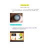
The eye has three layers: external, intermediate, and internal. External layer is a white, tough, and fibrous protective layer called the sclera. Light enters through the cornea, a transparent part of the sclera at the front. Intermediate layer is called the choroid, which absorbs stray light and contains blood vessels that nourish the eye. At the front, the choroid forms the iris, which contains the pupil. The iris adjusts the size of the pupil based on light condition which is called adaptation. Behind the iris the choroid thickens and forms the ciliary muscles, which is attached to the lens and focuses images on the retina. Internal layer is the retina, a thin layer of tissue that contains rods and cones. Rods are sensitive to light intensity and cones are sensitive to colours. Cones are more concentrated in the fovea centralis. They send sensory impulses to the brain via the optic nerve. The anterior chamber is at the front which is filled with aqueous humour and the posterior chamber is filled with vitreous humour. 88106366675
Focusing
Focusing on Distant Object:
Ciliary muscles relax
Tension on suspensory ligament
Tension pulls on lens
Lens flattens
Focus on far objects
Focusing on Near Object:
Ciliary muscles contract
Tension on suspensory ligament relaxes
Less pull on lens
Lens bulges
Focus on near objects
The ability of the lens to change shape in order to focus images clearly on the retina is a reflex called accommodation.1371600476250
Refraction
Light enters the eye, speed of light slows and light rays are bent. Process called refraction. Refraction occurs when light passes through the cornea. Remaining refraction occurs at the lens. When information reaches the brain, it’s processed so we can perceive the image correctly right side up and left to right.1285875866775
Optic Chiasm
Information you receive from each eye is communicated through the optic nerve to the brain. The brain processes this information into one image without gaps or blind spots. At optic chiasm, some axons from each eye cross over to the opposite side of the brain while others continue to their respective side of the brain. The crossing over ensures that each half of the occipital lobe receives the same image or part of the visual field as viewed by each eye. Data from the right half of each retina go to the right side of the occipital lobe and data from the left half of each retina go to the left side of the occipital lobe.
18811889525
Depth Perception
We take information from where an image falls on the retina. Due to the distance between the two eyes, images will not fall on the same spot on the retina for both eyes. When an object is placed closer, its image will fall on the inner portion of the retina, toward the nose. For objects placed farther, their image will fall on the outer portion of the retina, toward the ears. By comparing where an image falls on the retina, we can determine its distance from us.
Photoreception Pathway
Light
Cornea
Aqueous humour
Lens
Vitreous humour
Retina (cones and rods)
Optic nerve
Brain (occipital lobe)
The Retina (Rods & Cones)
The retina turns light energy into electrochemical impulses which are sent to the brain. Rods distinguish degrees of black and white and motion, are spread throughout the retina. Cones are colour-detecting, require intense light to stimulate them, are densely packed at the fovea centralis, allow us to perform high-acuity tasks (e.g. reading), three types of cones: red, blue, green.
How do photoreceptors relay visual information to the brain? Rods contain a light-absorbing pigment called rhodopsin, composed of retinal (vitamin A deritative) and the protein opsin. In dark, rods release an inhibitory neurotransmitter that inhibits nearby nerve cells. When rods absorb light, the rhodopsin splits into retinal and opsin, triggering a chain reaction that stops the release of inhibitory neurotransmitters, allowing transmission of neural impulses to the optic nerve. Cones process is similar, except the pigment is photopsin and only reacts to certain wavelengths of light. Opsin pigment absorbs light, the retinal molecule changes shape, initiates nerve impulse to be processed by occipital lobe of the cerebrum. Opsin pigment is bleached and cannot send signals, until retinal molecule reverts to original shape. -114299381000
Pigmented cells
closest to the choroid
specialized to form the tapetum in some animals
Rods and cones
the actual photoreceptors
located above the pigmented layer
You might expect the photoreceptors to be in the direct path of incoming light, but the rods and cones are covered by layers of transparent neurons.
Bipolar cells
activated by rods and cones
Ganglion cells
closest to the vitreous humour
Conditions
Glaucoma
Ducts that drain the aqueous humour are blocked; pressure ruptures blood vessels in eye and causes deterioration of the cells because lack of oxygen and nutrients.
Cataracts
Cloudy area on lens of eye caused by degeneration of protein structure of lens; prevents passing of light.
Macular degeneration
Affects people aged 50 and older. Caused by damage to macula, area surrounding fovea centralis, needed for sharp, central vision.
Cigarette smoking, obesity, and exposure to sunlight affect eyesight. Diabetic retinopathy, retinal detachment.
*Pages: 410-418
Lesson 3: Mechanoreception - The Ear
Key Terms
Sound Waves
Small fluctuation is air pressure resulting from sound, which causes particles around the source to vibrate and move; the auditory system detects these movements and the brain percieves them as sound.
Outer Ear
One of the three separate segments of the ear; consists of the pinna and the auditory canal.
Pinna
The outside flap of the ear; made of skin and cartilage and shaped in a way that enhances sound vibrations and focuses them into the ear.
Auditory Canal
Tube that conducts sound waves from the outer ear to the tympanum of the middle ear; amplifies sound waves, effectively making sounds louder.
Middle Ear
One of the three separate segments of the ear; begins at the tympanum and ends at two small openings in the wall of the inner ear called the round window and oval window.
Tympanum
Round, elastic structure within the middle ear that vibrates in response to sound waves.
Ossicles
Group of three small bones between the tympanum and oval window; transmits sound waves from the eardrum to the inner ear.
Oval Window
Membrane-covered opening in the wall of the inner ear; receives vibrations from the stapes.
Eustachian tube
Bony passage extending from the middle ear to the throat; plays a role in equalizing air pressure on both sides of the eardrum.
Inner ear
One of the three separate segments of the ear; consists of three components: semicircular canals, vestibule and cochlea.
Cochlea
One of the three main components of the inner ear; involved in hearing; within the cochlea, the mechanical energy of sound is converted into electrochemical impulses that are transmitted to the brain.
Organ of Corti
Organ of hearing found within the cochlea of the inner ear; contains hair cells that detect vibrations in the inner ear and transmit this information to the auditory nerve.
Basilar Membrane
One of the two parallel membranes that comprise the organ of Corti in the inner ear; lies along the base of the organ of Corti; attached to it are sensory mechanoreceptors known as hair cells.
Hair cells
Sensory mechanoreceptor attached to the basilar membrane.
Tectorial membrane
One of the two parallel membranes that comprise the organ of Corti; during transmission of sound waves, the basilar membrane vibrates, causing the sensory hairs to flex against the tectorial membrane.
Semicircular canals
One of the three main components of the inner ear; consists of three fluid filled loops, arranged in three different planes; contains mechanoreceptors that detect head and body rotation.
Rotational equilibrium
Balance required while rotating the head and body.
Gravitational equilibrium
Balance required while moving the head forward and backward.
Utricle
Saclike cavity in the vestibule of the inner ear; contains sensory receptors for gravitational equilibrium.
Saccule
Saclike cavity in the vestibule of the inner ear; contains sensory receptors for gravitational equilibrium.
Otoliths
Calcium carbonate granule associated with sensory receptors for detecting movement of the head; in vertebrates, located in the utricle and saccule on the vestibule of the inner ear.
Three major regions: Outer Ear, Middle Ear & Inner Ear.
Pathway of Sound
pinna ? auditory canal ? tympanum (eardrum) ? ossicles ? oval window ? cochlea (organ of Corti) ? auditory nerve ? brain (temporal lobe)
Outer ear
Consists of the pinna and auditory canal. Sound waves collected by the pinna are directed down the auditory canal to the tympanic membrane. Ear wax is produced to moisten the passage and tympanic membrane for flexibility. Hairs prevents dust and foreign materials from proceeding. The auditory canal ends at the tympanum/tympanic membrane. Sound waves cause it to vibrate, which converts sound energy to mechanical energy.
Middle ear
Consists of the tympanum, ossicles and Eustachian tube. Tympanic membrane is connected to the ossicles, which conduct and amplify mechanical vibrations to the oval window. Middle ear is connected to the throat by the Eustachian tube which allows air pressure to equalize.
Inner ear
Composed of two organs: one for hearing and one for balance. Hearing: oval window, cochlea, round window and auditory nerve. Balance: utricle, saccule, and semicircular canals. Inner ear converts mechanical stimulation to a nerve impulse, and mechanical stimulation into information on balance to be interpreted by the brain. Vibrating ossicles cause the oval window to vibrate. This causes pressure waves in the fluid-filled canals of the cochlea. The waves travel to the round window that moves to equalize pressure.
Cochlea - Organ of Corti297656338100
Cochlea converts mechanical energy into electrochemical impulses that are transmitted to the temporal lobe of the brain. The middle chamber of the Cochlea contains the organ of Corti. The base of the organ of corti is the basilar membrane which mechanoreceptors (hair cells) are attached. Hair cells have thin projections called stereocilia which stick out the top of the cells. The far ends are embedded within the tectorial membrane. Pressure waves make the basilar membrane move up and down, causing the stereocilia of the hair cells to bend against the tectorial membrane. The hair cells, which synapse with the nerve fibers of the auditory canal, sense the bending and relay the message to the nerves.
Sound
Frequency is the number of vibrations in a given unit of time; determines pitch, which is the degree of highness or lowness of a sound. Amplitude refers to the distance from the rest position to the crest of the sound wave; as the amplitude increases, the sound becomes louder.
The basilar membrane closest to the oval window (where frequency is 20,000 Hz) is narrow and stiff; high frequency sound waves. The basilar membrane by the apex or tip of the cochlea (where frequency is 20 Hz) is wide and flexible; low frequency sound waves. Humans hear between 20 and 20 000Hz.
Hearing Loss
Deafness is one of two main types: conduction deafness or perception deafness. Perception (nerve) deafness: any of the structures of the organ of Corti have been damaged (e.g. Hair cells, the sensory neurons, the basilar membrane, the tectorial membrane, or the auditory nerve). Conduction deafness, structures like the tympanum, any of the ossicles, or the connection between the ossicles and the oval window have been damaged.
Perception of Sound
Sensory neurons send information through the auditory nerve, to the brain stem, thalamus, and the temporal lobe for processing. Depending on what sensory neurons are stimulated, the brain can perceive the frequency and amplitude of sound. Brain can also detect the location where the sound came from.
Balance and Coordination
Three structures: semicircular canals, utricle and saccule. Semicircular canals contain mechanoreceptors which detect rotation (rotational equilibrium). Arranged in three different planes, the base ends in a bulge. Inside the bulge, stereocilia of the hair cells stick into a jelly-like covering called a cupula. Rotation causes the fluid to move and bend the stereocilia causing hair cells to send information to the brain. Gravitational equilibrium is the balance required when moving your head forward or backwards.The utricle and saccule contain calcium carbonate granules called otoliths, which lie in a capula over a layer of hair cells. When you move your head, gravity pulls on the otoliths, bending the hair cells and sending a neural impulse to the brain indicating the position of the head. Proprioceptors are found in muscles, tendons and joints and send information about body position to the brain.
*Pages: 419-425
Lesson 4: The Structure and Organization of the Nervous System
Homeostasis
Nervous system
Central nervous system
Peripheral nervous system
The nervous system is two systems: Central Nervous System (CNS) and Peripheral Nervous System (PNS).
Central Nervous System: Brain and spinal cord.
Peripheral Nervous System: Everything else, carries sensory information to the CNS and send information from the CNS to the motor pathways.
Peripheral Nervous System
Information sent from CNS to muscles and glands controls the body consciously and unconsciously.
Somatic Nervous System: Controls voluntary movement.
Autonomic Nervous System: Controls involuntary movement.
Sympathetic Nervous System: Fight-or-Flight.
Parasympathetic Nervous System: Rest and Digest.
Sympathetic Nervous System: Triggered during stressful situations to mobilize energy. Uses epinephrine to control organ responses.
Parasympathetic Nervous System: Triggered during relaxed states to converse energy. Uses acetylcholine to control organ responses.
Sympathetic Nervous System
Parasympathetic Nervous System
Increased heart rate
Inhibits tears and salivation
Dilates pupils
Liver releases glucose
Inhibits digest
Inhibits defecation
Inhibits urination
Decreased heart rate
Stimulates tears and salivation
Constricts pupils
Gallbladder release bile (more digestion)
Stimulates digestion
Stimulates defecation
Stimulates urination
Homeostasis is the maintenance of a nearly constant internal environment that fluctuates at an ideal point. Function of the nervous system is to collect information, analyze (sort, integrate, interpret) the information and to initiate an appropriate response to maintain a state of homeostasis.
Central Nervous System
The Brain
It’s protected by a bony exterior called the skull. Beneath the skull are three layers of tough, elastic connective tissue called meninges.
Blood from the circulatory system doesn’t enter the brain and spinal cord directly because of the blood-brain barrier. This selective barrier protects the brain from toxins and bacteria but allows oxygen, glucose, and other lipid-soluble substances to pass.
Cerebrospinal fluid (CSF) is a fluid that surrounds the brain and the spinal cord. It protects the central nervous system from injury. It is produced constantly throughout the day and transports nutrients, white blood cells, and hormones.
Forebrain, Midbrain, and Hindbrain
Hindbrain consists of a wrinkled ball at the base of the brain called the cerebellum and the upper part of the brainstem, the medulla oblongata and the pons. Pons is a relay centre for the brain, it connects the cerebrum to the cerebellum. Medulla oblongata controls involuntary movements such as breathing, heart rate, blood pressure, and swallowing. Cerebellum controls body movement and balance.
Midbrain is above pons and controls eye movement muscles and other skeletal muscles.
Forebrain consists of the cerebrum, thalamus, and hypothalamus. Thalamus is another relay centre for the brain, connects the neurons in the PNS to the brain. Hypothalamus maintains homeostasis (body temperature, blood volume, water balance, blood pressure, and thirst), regulates emotions, and controls the pituitary gland. Cerebrum is split into two hemispheres, has centres for intellect, memory, consciousness, and language.
Cerebrum
The cerebral cortex is the outer covering of grey matter is responsible for language, memory, personality, conscious thought and other activities associated with thinking and feeling. Divided into left and right hemispheres that communicate through the corpus callosum. The left hemisphere is responsible for language ability, and the right hemisphere controls reasoning, spatial abilities, and visual recognition.
The cerebrum has four major lobes: frontal lobe, parietal lobe, occipital lobe, and temporal lobe.
The frontal lobe is responsible for intellect, learning, memory, personality, emotions, creativity, and voluntary motor movement.
The parietal lobe is responsible for processing sensory information (touch and taste).
The occipital lobe is responsible mainly for processing visual information.
The temporal lobe is responsible mainly for processing auditory information and contains Wernicke’s area.
The Spinal Cord
Spinal cord is the communication between the brain and the peripheral nervous system. It’s protected by the backbone and the cerebrospinal fluid. Contains white and grey matter. The outer white matter is myelinated nerve fibres. The inner white matter is unmyelinated neurons, cell bodies and dendrites.
The spinal cord has two special types of nerves.
Sensory nerves (dorsal root) communicate messages from the body to the brain for interpretation.
Motor nerves (ventral root) communicate messages from the brain to effectors (muscles) that initiate responses.
The dorsal root ganglion is composed of cell bodies of sensory neurons.
MRI and PET Scan
PET scan looks at functions of the brain. Radioactive substance is tagged to glucose and ingested, shows various colours and brightness according to activity levels. MRI looks at the structure. Uses magenets and radio signals to produce clear images of organs.
Pages: 366-367, 385-395