Transcript
By: Jessica Davis
Ms. Silva – Microbiology
Ebola Hemorrhagic
Fever
Ebola virus is a member of the Filoviridae viruses, a family of viruses that cause severe and frequently fatal hemorrhagic fevers
Single stranded ribonucleic acid, each virus is a thread enveloped by protein-coated RNA
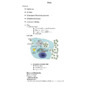
Viral Structure of Ebola
Distinct characteristic “6” shape
ss, negative sense RNA
At least a 30% genetic variation between the different Ebola strains
Five different strains of the Ebola virus have been identified in Africa and America, which include:
Reston (initially from the Philippines)
Zaire (most lethal)
Sudan
Ivory Coast (rarest)
Uganda (Bundibugyo ebolavirus)
Ebola Zaire (EBO-Z): a < 90% death rate
Ebola Sudan (EBO-Z): lower death rate
Ebola Reston: a 71% death rate
Ebola Cote d’ Ivory: no reported fatalities
Bundibugyo ebolavirus: a 71% death rate
All strains of Ebola are classified as Biosafety Level 4, meaning Hazmat suits, multiple airlocks, ultraviolet light rooms. Workers must be cleared to handle BSL4.
Strains
These are examples of the posters posted throughout villages in Africa in hopes minimizing the spread of Ebola
The main and easiest method of transmission in Ebola is through bodily fluids (blood, secretions).
Infections contracted through direct contact more often result in death
Animals such as fruit bats (Zaire strain) and arthropods (mainly to humans) via bites
Handling infected animals can also lead to infection of Ebola; scientists observed that it may be transferred aerobically from dead animal carcasses to humans
Monkeys mainly transfer Ebola between themselves via airborne particles
Transmission
Internal bleeding is caused by Ebola’s coagulopathy ability. This describes a dysfunction in the host blood clotting system.
When infected, host macrophages begin to express Tissue Factor (TF). TF attracts clotting molecules from the blood, leaving the rest of the body susceptible.
Small holes in the capillaries are then cut by Ebola. Without clotting factors, the host bleeds continuously, dying of what some have called “a million cuts.”
Coagulopathy
Incubation periods can be anywhere from 2-21 days.
Common symptoms include: sudden onset of fever, headaches, sore throat, muscle pains, and intense weakness.
More intense symptoms include: maculopapular rash, kidney/liver dysfunction.
Possible internal/external bleeding.
Debilitating effects on endothelial cells along with shock produce the most fatalities.
Symptomology
1999: BBC researchers, led by Dr. Maurice Iwu, investigated the garcin kola plant, typically Eastern in Western Africa. Medicine men in those areas had long been using it and introduced it to the researchers. In a lab setting, the plant has been shown to inhibit Ebola multiplication.
2001: Mice injected subcutaneously with Ebola did not become sick, but mounted an immune response. Serum from these mice were used to treat new mice before or after Ebola injection. All of the mice treated with serum survived.
2004: Scientists first investigated the idea of “prime boosting” in monkeys. First, the host was treated with noninfectious genetic material from a pathogen. Weeks later, the attenuated form of a virus was injected, eliciting a faster immune response.
This 2nd step was shown to cause a faster (yet weaker) immune response, which is key in trying to protect a community from a rampant outbreak.
The scientists used this boosting effect with an EBO-Z strain that killed its victim. All of the monkeys were protected, even at high doses.
Cures/Vaccines
Cures/Vaccines
Recently it has been discovered that there are two cellular enzymes Ebola virus must have to reproduce. When those enzymes are blocked, the virus loses most of its infectivity
Scientists has been testing various treatments that block these enzymes in primates and have been fairly successful, but more testing is still being done
In the picture: The enzyme blockers surrounding the virus blocking enzymes and in doing so preventing the Ebola from reproducing