Endocrine System
Endocrine Vs Nervous System
The endocrine system acts with nervous
system to coordinate
the body's activities.
Both systems enable cells to communicate with others by using
chemical messengers.
The endocrine system uses chemical messengers called hormones that
are transported by the circulatory system (blood). They act on
target cells that may be anywhere in the body.
The endocrine system is slower than the nervous system because
hormones must travel through the circulatory system to reach their
target.
Target cells have receptors that
are specific to the signaling molecules. The binding of
hormones to the receptors on or within the target cell produces a
response by the target cell.

The chemical messengers used by the nervous system are neurotransmitters.
Neurotransmitters travel across a narrow space (the synaptic
cleft) and bind to receptors on the target cell.
The nervous system conducts signals much quicker than the
endocrine system.
Endocrine Vs Exocrine glands
Endocrine glands do not have ducts. Exocrine glands have ducts
that carry their secretions to specific locations.
Two Kinds of Hormones
Peptide Hormones
Peptide hormones are composed of amino acids.
A peptide hormone binds to a cell-surface receptor, it does not
enter the cell.
The resulting complex activates an enzyme that catalyzes the
synthesis of cyclic AMP from ATP. Cyclic AMP activates other enzymes
that are inactive.

Cyclic AMP is a second messenger; the hormone is the first
messenger. Other second messengers have been discovered.
Steroid Hormones
Steroid hormones enter the cell and bind to receptors in the
cytoplasm.
The hormone-receptor complex enters the nucleus where it binds
with chromatin and activates specific genes. Genes (DNA) contain
information to produce protein as diagrammed below. When genes are
active, protein is produced.


Steroid hormones act more slowly than peptide hormones because of
the time required to produce new proteins as opposed to activating
proteins that are already present.
Hypothalamus
The hypothalamus is
part of the brain. It maintains homeostasis (constant internal
conditions) by regulating the internal environment (examples: heart
rate, body temperature, water balance, and the secretions of the
pituitary gland).
Pituitary Gland
The pituitary contains two lobes. Hormones released by the
posterior lobe are synthesized by neurons in the hypothalamus.
Unlike the posterior lobe, the anterior lobe produces the hormones
that it releases.
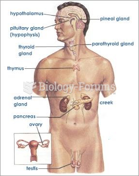
Refer to the diagram below as you read about the hypothalamus,
pituitary, and each of the glands they control.

Posterior pituitary
The posterior pituitary contains axons of neurons that extend
from the hypothalamus. Hormones are stored in and released from axon
endings in the posterior lobe of the pituitary.
Oxytocin
Oxytocin stimulates the uterine contractions of labor that are
needed to move the child out through the birth canal.
The hormone stimulates the release of milk from the mammary
glands by causing surrounding cells to contract. After birth,
stimulation of the breast by the infant feeding stimulates the
posterior pituitary to produce oxyticin.
Antidiuretic Hormone (ADH)
Antidiuretic hormone increases the permeability of the distal
convoluted tubule and collecting duct of the kidney nephron
resulting in less water in the urine. The urine becomes more
concentrated as water is conserved.
The secretion of ADH is controlled by a negative feedback
mechanism as follows:
concentrated blood (too little water) ®
hypothalamus ®
ADH ®
kidney ®
reabsorbs water, makes blood more dilute
Below: Within the kidney, fluid and dissolved substances are
filtered from the blood and pass through tubules where some of the
water and dissolved substances are reabsorbed. The remaining liquid
and wastes form urine. Details of this process are discussed in the chapter
on the excretory system.

The presence of too much blood in the circulatory system
stimulates the heart to produce a hormone called atrial
natriuretic factor (ANF).
This hormone inhibits the release of ADH by the posterior pituitary
causing the kidneys to excrete excess water.
Alcohol inhibits the release of ADH, causing the kidneys to
produce dilute urine.
Control of the Anterior Pituitary
The hypothalamus produces hormones that travel in blood vessels
to the anterior pituitary, stimulating it to produce other hormones.
The hormones produced by the hypothalamus are called hypothalamic-releasing
hormones.

The anterior pituitary produces at least six different hormones.
Each one is produced in response to a specific
hypothalamic-releasing hormone.
The blood vessel that carries hypothalamic-releasing hormones
from the hypothalamus to the pituitary is called a portal
vein because
it connects two capillary beds. One capillary bed is in the
hypothalamus and the other is in the anterior pituitary.
Release-inhibiting hormones produced by the hypothalamus inhibit
the pituitary from secreting its hormones.

Example
The pituitary is stimulated to release growth hormone (GH) by
growth hromone releasing hormone (GHRH) produced in the
hypothalamus. It is inhibited from releasing growth hormone by
growth hormone release-inhibiting hormone(GHRIH), also produced by
the hypothalamus.

Six different hormones produced by the anterior lobe will be
studied here. Three of these have direct effects on the body, the
other three control other glands.

Anterior Pituitary Hormones that Directly Affect the Body
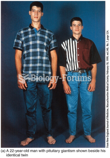
Growth Hormone (GH or Somatotropic Hormone)
Growth hormone stimulates body cells to grow. If too little
hormone is produced, pituitary dwarfism results. The secretion
of too much hormone results in a pituitary giant.
Acromegaly is a genetic disease in which growth hormone is
produced throughout a persons lifetime.
Prolactin
Prolactin is produced in quantity after childbirth.
It stimulates the development of the mammary glands and the
production of milk.
It is also involved in the metabolism of fats and carbohydrates.
Melanocyte-Stimulating Hormone (MSH)
This hormone causes skin color changes in some fishes,
amphibians, and reptiles.
In humans, it stimulates the melanocytes to synthesize melanin.
Anterior pituitary hormones that regulate other glands
The pituitary also controls other glands and is often referred to
as the "master gland".
Three kinds of pituitary hormones that regulate other glands are
discussed below. The glands that they regulate will be discussed in
the following section.
Thyroid Stimulating Hormone (TSH) ® thyroid ®
thyroxin
Adrenocorticotropic Hormone (ACTH) ®
adrenal cortex ®
cortisol
Gonadotropic Hormones (FSH and LH) ®
ovaries and testes ®
sex hormones; controls gamete production
Negative Feedback Inhibition
Hormone secretions by glands that are under the control of the
hypothalamus are controlled by negative
feedback. When
the hormone levels are high, they inhibit the hypothalamus and
anterior pituitary, resulting in a decline in their levels.

The thyroid produces thyroxin (also
called T4 because
it contains 4 iodine atoms) and triiodothyronine (also
called T3 because
it contains 3 iodine atoms).
Both T4 and
T3 have
similar effects on target cells. In most target tissues, T4 is
converted to T3. They influence metabolic rate, growth,
and development.
Thyroxin production is regulated by a negative feedback mechanism
in which it inhibits the hypothalamus from stimulating the thyroid.

Hypothyroidism occurs
when the thyroids produce too little hormone. In adults, it results
in lethargy and weight gain. In infants, it causes cretinism, which
is characterized by dwarfism, mental retardation, and lack of sexual
maturity. Administering thyroid hormones treats these affects.
Too much T3 and
T4 (hyperthyroidism)
increases heart rate and blood pressure, and causes weight loss.
Iodine is needed to manufacture thyroid hormones. A deficiency in
iodine prevents the synthesis of thyroid hormones which, in turn,
results in an excess of thyroid stimulating hormone being produced
by the anterior pituitary. A goiter results
when constant stimulation of the thyroid causes it to enlarge.
Calcitonin
The thyroid gland also secretes calcitonin,
which stimulates calcium deposition in the bones. This is the
opposite of the action of parathyroid hormone (see below).

Calcitonin production is
not regulated by
the anterior pituitary. It's secretion is stimulated by high calcium
levels in the blood.
Parathyroid glands
The parathyroid glands are 4 small glands embedded in posterior
surface of the thyroid gland.
They secrete parathyroid
hormone (PTH), which increases blood levels of Ca++.
Bone tissue acts as a storage reservoir for calcium; PTH
stimulates the removal of calcium from the bone to increase levels
in the blood.
PTH also increases the kidney’s reabsorption of Ca++ so
that less is lost in urine and it activates vitamin D which enhances
Ca++ absorption
from food in the gut.
Secretion is regulated by the Ca++ level
in the blood, (not hypothalamic or pituitary hormones).
The outer layer of an adrenal gland is the adrenal cortex.
It produces three kinds of steroid hormones. These are glucocorticoids, mineralocorticoids,
and small amounts of sex hormones. The major glucocorticoid is cortisol and
the major mineralocorticoid is aldosterone.
Cortisol (A Glucocorticoid)
Glucocorticoids are produced in response to stress.
Cortisol raises the level of glucose in the blood by stimulating
the liver to produce glucose from stored non-carbohydrate sources
such as proteins and lipids and
to release it into the blood.
Cortisol reduces swelling by inhibiting the immune system.
Swelling of tissues due to injury or infection is discussed in the chapter
on the immune system. The drug prednisone, derived from
cortisol, is used to treat inflammation.
Negative feedback control of cortisol level is diagrammed below.

Aldosterone (A Mineralocorticoid)
Aldosterone secretion is not under the control of the anterior
pituitary.
It acts primarily on the kidney to promote absorption of sodium
and excretion of potassium.
Increased sodium levels contributes to the retention of water and
thus increased blood volume. In the absence of aldosterone,
sodium is excreted and the lower sodium levels result in decreased
blood volume and lower blood pressure.
The presence of too much blood in the circulatory system
stimulates the heart to produce atrial
natriuretic factor. This hormone inhibits the release of
aldosterone by the adrenal cortex and ADH by the posterior pituitary
causing the kidneys to excrete excess water. The loss of water and
sodium contribute to lowering the blood volume.

The adrenal medulla is composed of modified neurons that secrete
epinephrine and norepinephrine (adrenaline and noradrenaline) under
conditions of stress.
These hormones are released in response to a variety of stresses
and stimulate the fight- or- flight response of the sympathetic
nervous system. It results in a faster heart rate, faster blood
flow, and dilated airways to facilitate oxygen flow to the lungs. In
addition, the level of glucose in the blood is increased to make
energy more available.
Their secretion is controlled by brain centers (including
hypothalamus) via sympathetic nerves, not by pituitary hormones.
LH and FSH from
the anterior pituitary stimulate the gonads (ovaries and testes).
LH stimulates the testes to produce several kinds of steroid
hormones called androgens.
One of these androgens is testosterone,
the main sex hormone in males.
LH stimulates the ovaries produce estrogen and progesterone,
the female sex hormones.
Sex hormones are responsible for the development of secondary
sex characteristics, which develop at puberty. Some examples
of secondary sex characteristics in males are deepening of the voice
(due to a large larynx), growth of facial hair, and muscle
development. Some secondary sex characteristics in females are
development of the breasts and broadening of the pelvis. Both sexes
show increased activity of sweat glands and sebaceous glands (oil
glands in the skin), and growth of pubic and axillary (armpit) hair.
FSH controls gamete (egg or sperm) production.

The pancreas is a digestive gland that secretes digestive enzymes
into the duodenum through the pancreatic duct.
The islets of
Langerhans are
groups of cells within the pancreas that secrete insulin and glucagon.
The islets are endocrine glands because they are ductless; the
circulatory system carries their hormones to target cells.
Insulin
Insulin promotes the removal of glucose from the blood for
storage as glycogen (muscle, liver), fats (fat cells), and protein.
It promotes the buildup of fats and proteins and inhibits their
use as an energy source.
Glucagon
Glucagon is produced in the islets of Langerhans but by different
cells than those that produce insulin.
The effects of glucagon are opposite those of insulin. It
raises the level of glucose in the blood.
It is normally secreted between meals to maintain the
concentration of glucose in the blood.

Diabetes Mellitus
Diabetes mellitus is a disease in which glucose is not
sufficiently metabolized. This results in high glucose levels in
blood and glucose in the urine.
Cells can starve because glucose is not being metabolized.
Type I
Type I diabetes is also called "juvenile-onset diabetes" or
"insulin-dependent diabetes" because the symptoms usually appear
during childhood and insulin injections are necessary to treat it.
It usually occurs after a viral infection triggers an immune
response that results in the body destroying its own
insulin-producing cells.
Because the disease is caused by a lack of insulin, it can be
treated with insulin injections.
Type II
Type II diabetes is more common than type I.
Type II diabetes is caused by a deficiency in insulin production
or by changes in insulin receptors on the target cells. In either
case, blood glucose level may be high because cells do not receive
the message to metabolize glucose.
This form of diabetes usually becomes noticeable in middle age.
It is treated with a low fat, low sugar diet, regular exercise,
weight control. Another treatment is oral medications that make the
cells more sensitive to the effects of insulin or that stimulate
more insulin production.
Thymus Gland
The thymus grows during childhood but gradually decreases in size
after puberty.
Lymphocytes that
have passed through the thymus are transformed into T
cells.
Lymphocytes are white blood cells that function to fight
infection. There are two kinds of lymphocytes: B cells and T
cells. T cells participate in identifying and destroying
body cells that are infected.
Thymus hormones called thymosins stimulate
the development and differentiation of T lymphocytes. They play a
role in regulating the immune system by stimulating other kinds of
immune cells as well.

Pineal Gland
Fish and Amphibians
The pineal gland of fish and amphibians is located near the skin
and functions to detect light.
Birds and Mammals
In birds, it is located on the brain but still receives direct
light stimulus through the skull.
In mammals, it is located within the brain and therefore cannot
receive light stimulation directly. Light from the eyes stimulates
the gland via the optic nerve.
Melatonin is produced when the pineal gland is in the dark.
During the winter, nights are longer and as a result the level of
melatonin in the blood is higher. The level of melatonin in
the blood therefore varies with season and can be used to help
animals time events such as when to breed, nest, migrate, etc.
These annual cycles are called circannual
rhythms. Melatonin may also participate in
producing 24-hour cycles called circadian
rhythms.
In humans, the gland may be involved in sexual development.
Flashcards
The next five pages contain flashcards that can be used to learn
the glands and their secretions. Use scissors to cut out the
flashcards.
1) Eleven of the cards contain gland names written on one side.
Write the name of the secretion on the other side. Go through these
cards by viewing the gland name and trying to identify the
secretion. Then, go through the cards by viewing the secretion name
and trying to identify the gland name. Continue going through all of
the cards until you have learned all of the glands and their
secretions.
2) Twenty-three cards have secretions written on one side. Write
the following information on the other side:
-The name of
the gland that produces the hormone
-How the
hormone affects the body
-How production
of the hormone is controlled
Go through these
cards by viewing the secretion name and trying to state the effect
of the hormone and then telling how the hormone is controlled.
Continue going through the cards until you have learned how the
hormones affect the body and how the hormones are controlled.
|
Glands
adrenal
cortex
adrenal
medulla
anterior
pituitary
ovaries
testes
pancreas
parathyroid
pineal
posterior
pituitary
thymus
thyroid
|
Secretions
adrenocorticotropic hormone
aldosterone
antidiuretic hormone
calcitonin
cortisol
epinephrine, norepinephrine
estrogen
FSH
(follicle stimulating hormone)
glucagon
gonadotropic hormones (FSH, LH)
growth
hormone
insulin
LH
(leutinizing hormone)
melatonin
oxytocin
parathyroid
hormone
prolactin
progesterone
testosterone
thymosins
thyroid
stimulating hormone
thyroxin
triiodothyronine
|
|
 Quick Reply