|
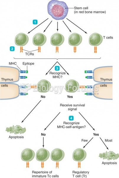
A normal immune response begins in a peripheral tissue wherein an ‘offending’ antigen is endocytosed by a dendritic cell and thence transferred to the regional lymph node. Here an immune response proceeds via the germinal centre reaction in lymphoid follicles wherein there occurs activation of T and B lymphocytes with return to the affected tissue of activated T cells and specific antibody to dispose of the inciting antigen, so terminating the response. Very likely, autoimmune responses proceed in the same way,30 but with important provisos. Thus, for an autoimmune response to proceed, there needs to be coexisting inflammation such that antigen-presenting cells (APCs) are appropriately activated, natural immune tolerance must be in some way compromised and, finally, the response does not terminate because auto-antigen cannot be eliminated. Of course, there are many uncertainties in this scenario including the source and degree of the coexisting inflammation, the range of stimuli that activate Toll-like receptors (TLR) on a APC, and whether the initial stimulus is provided by spillage of native auto-antigen, by an apoptotic fragment, or by an auto-antigenic mimic.31 Maintenance of the reaction in the regional lymph node is the next step. This depends on whether low-prevalence auto-antigen-responsive T cells, as escapees of central tolerance in the thymus, happen to be en passage through the node, whether various cytokine–chemokine signalling pathways are conducive, whether processes of apoptosis are intact and, overall, the nature of the deficits in immune tolerance. Although tolerance was predicted by Medawar32 in 1960 as likely to be comprehended ‘within a few years’, this extraordinarily complex process comprises as many as 24 ‘overrides’ or ‘checkpoints’ that stand in the way before a potentially auto-reactive immunocyte attains pathogenic reactivity.33 Finally, the direct effector processes aimed towards eliminating a foreign antigen or, in the case of autoimmunity, an auto-antigen, comprise activated CD4+ cytokine- releasing T cells, CD8+ cytolytic T cells (CTLs) and autoantibody-producing B cells, with down-stream recruitment of many effector elements including invariant NKT or NK cells, macrophages, mast cells, Fc receptors, complement and others. Evidence is accruing particularly for effector activity of CD8+ CTLs in autoimmune injury, for example in type 1 diabetes,34 and in primary biliary cirrhosis (PBC),35 which would be consistent with the capacity for display on the cell surface by MHC Class I molecules of peptides derived from intracellular autoantigens.
Until recently, autoimmunity was usually considered essentially as a pathology of the adaptive immune response. However, it is now recognized that some autologous molecules are capable per se of activating elements including TLR of the innate immune system. While in these situations T cells usually participate, even these are dispensable according to a mouse model of lupus driven by the B-cell stimulatory cytokine BAFF and elements of innate immunity.
|
 Quick Reply