|
Cerebral Cortex
The cerebral cortex is 1.5 to 4.0 mm thick and contains about 50 billion neurons plus nerve processes and supporting glial cells. In all but a few regions it is characterized by a laminated appearance. Perikarya generally are organized into five layers. Starting at the periphery of the cerebral cortex, the general organization of neurons is molecular layer (I), external granular layer (II), external pyramidal layer (III), internal granular layer (IV), internal pyramidal layer (V), and multiform layer (VI).
The molecular layer (I) is a largely perikaryon-free zone just below the surface of the cortex. Neurons of similar type tend to occupy the same layer in the cerebral cortex, although each cellular layer is composed of several different cell types. For convenience of description, these neurons often are placed in two major groups: pyramidal cells and stellate or nonpyramidal cells. The perikarya of pyramidal cells are pyramidal in shape and have large apical dendrites that usually are oriented toward the surface of the cerebral cortex and enter the overlying layers; the single axons enter the subcortical white matter. They are found in layers II, III, V, and, to a lesser extent, layer VI. Very large pyramid-shaped neurons (Betz cells) are present in the internal pyramidal layer (V) of the frontal lobe. Stellate (nonpyramidal) cells lack the pyramid-shaped perikarya and the large apical dendrite. They occur in all layers of the cerebral cortex but are concentrated in the internal granular layer (IV). Impulses entering the cortex are relayed primarily to stellate cells and then transmitted to pyramidal cells in the various layers by the vertical axons of the stellate cells. Axons of pyramidal cells generally leave the cortex and extend to other regions of the brain and spinal cord.
The cerebral cortex functions in vision, hearing, speech, voluntary motor activities, and learning.
Cerebellar Cortex
Three layers characterize the cerebellar cortex: an outer molecular layer, a middle Purkinje cell layer, and an inner granule cell layer.
The molecular layer is mainly a synaptic area with relatively few nerve cell bodies. It consists primarily of unmyelinated axons from granule cells, the axons running parallel to the cortical surface. It also contains large dendrites of the underlying Purkinje cells and in its superficial portion contains small-scattered neurons called stellate cells. Other small neurons located deep in this layer and adjacent to Purkinje cells are called basket cells.
The Purkinje cell layer is formed by the cell bodies of Purkinje cells - large, pear-shaped neurons aligned in a single row and characterized by large branching dendrites that lie in the molecular layer. They represent Golgi type I neurons and number about 15 million. Three-dimensionally, the large dendritic trees occupy a narrow plane, reminiscent of fan coral, and are so arranged that each dendritic tree is parallel to its neighbor. A single small axon from the Purkinje cell passes through the granule cell layer and synapses with neurons in the central cerebellar area.
The granule cell layer consists of numerous closely packed, small neurons whose axons enter the molecular layer to synapse with dendrites of Purkinje cells. The granule cells represent Golgi type II neurons. They have small, round nuclei with coarse chromatin patterns and only scant cytoplasm; dendrites are short and clawlike. The axons enter the molecular layer, bifurcate, and run parallel to the surface but perpendicular to the wide plane of the Purkinje dendritic tree. The axons of granule cells synapse with about 450 Purkinje cells in a relationship similar to that of wires coursing along telephone poles. Axons of granule cells also synapse with stellate and basket neurons in the molecular layer. Another type of small neuron, the Golgi cell, is found in the outer zone of the granule cell layer.
Two types of afferent nerve fibers enter the cerebellar cortex from outer regions of the central nervous system. These are the mossy fibers, which synapse with granule cells, and the climbing fibers, which enter the molecular layer and wind about the dendrites of Purkinje cells.
The cerebellum functions primarily in the modulation of skeletal muscle activity such as the coordination of limb movement and balance.
The gray matter of the spinal cord, cerebrum, and cerebellum consists of a complex, highly ordered meshwork of dendritic, axonal, and glial processes that envelop the perikarya of associated neurons. This network, called the neuropil, provides a vast area for synaptic contact and interaction between nerve processes and forms an organizing framework. It is important for coordinating activities in the central nervous system.
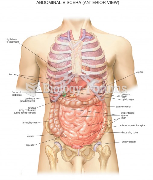
Dorsal View of the Brain (see attachment)
|


 Quick Reply