Transcript
Assignment 4: Investigating the Genetic Basis of Disorders
[30 PTS TOTAL]
Overview
The objectives of this activity are to learn more about DNA sequencing, and how changes to DNA, such as mutations, can influence protein structure and function. In Part A you will investigate how single nucleotide substitutions in the insulin-encoding gene impact protein formation. For this part, you will use the BLAST program to determine the location of nucleotide substitutions. In Part B you will become an “expert” on a genetic disorder of your choosing, and will respond to questions about how changes to particular genes impact the expression of the disorder.
Please turn in your answers to the below questions on Canvas by 9:00 am on Thursday, Oct. 13. This assignment is worth 30 points (over half the weight of an exam).
Part A. Which Insulin Mutations May Result in Disease?
Now that it's possible for individuals to have their whole genomes sequenced, doctors can use DNA sequence information to diagnose diseases and identify new treatments. One gene of great interest to medical researchers is the human insulin gene. The protein insulin is produced and secreted into the bloodstream by pancreatic ? cells as a key regulator of blood glucose levels. Recent work in medical genetics has demonstrated that severe cases of juvenile (type 1) diabetes can be caused by point mutations that alter the insulin coding sequence. Because these point mutations arise through single nucleotide substitutions, they are classified as single nucleotide polymorphisms (SNPs). Because SNPs are also used to trace the inheritance of genetic diseases such as diabetes, a large number of SNPs have been identified in the human insulin gene. But not all SNPs in the insulin gene result in diabetes in the patient. Suppose you are a medical geneticist presented with four patients, all of whom have a SNP in their insulin gene. It is your job to identify each SNP and figure out which patients are at risk of disease and which ones are not.
Section I. Using BLAST to identify SNPs
In the 1990s, scholars at the National Institute of Health designed a now-widely used bioinformatics program called BLAST (Basic Local Alignment Search Tool). This program compares unknown nucleotide or amino acid sequences to known sequences in online databases. It also can be used to align two or more closely related sequences to identify the locations of mutations.
Here, you will use BLAST to identify the locations and base substitutions of SNPs by aligning each patient's sequence with a reference sequence. In this case, the reference sequence is the cDNA sequence of the wild-type human insulin gene. cDNA (complementary DNA) is a piece of double-stranded DNA that contains only that portion of a gene that is translated (introns are not included) and is widely used to compare the coding regions of genes.
Below are five cDNA sequences of the human insulin gene: the wild-type sequence and sequences from four patients, each containing a single SNP.
Reference cDNA sequence (wild-type human insulin)
ATGGCCCTGTGGATGCGCCTCCTGCCCCTGCTGGCGCTGCTGGCCCTCTGGGGACCTGACCCAGCCGCAGCCTTTGTGAACCAACACCTGTGCGGCTCACACCTGGTGGAAGCTCTCTACCTAGTGTGCGGGGAACGAGGCTTCTTCTACACACCCAAGACCCGCCGGGAGGCAGAGGACCTGCAGGTGGGGCAGGTGGAGCTGGGCGGGGGCCCTGGTGCAGGCAGCCTGCAGCCCTTGGCCCTGGAGGGGTCCCTGCAGAAGCGTGGCATTGTGGAACAATGCTGTACCAGCATCTGCTCCCTCTACCAGCTGGAGAACTACTGCAAC
Patient 1 sequence
ATGGCCCTGTGGATGCGCCTCCTGCCCCTGCTGGCGCTGCTGGCCCTCTGGGGACCTGACCCAGCCGCAGCCTTTGTGAACCAACACCTGTGCGGCTCACACCTGGTGGAAGCTCTCTACCTAGGGTGCGGGGAACGAGGCTTCTTCTACACACCCAAGACCCGCCGGGAGGCAGAGGACCTGCAGGTGGGGCAGGTGGAGCTGGGCGGGGGCCCTGGTGCAGGCAGCCTGCAGCCCTTGGCCCTGGAGGGGTCCCTGCAGAAGCGTGGCATTGTGGAACAATGCTGTACCAGCATCTGCTCCCTCTACCAGCTGGAGAACTACTGCAAC
Patient 2 sequence
ATGGCCCTGTGGATGCGCCTCCTGCCCCTGCTGGCGCTGCTGGCCCTCTGGGGACCTGACCCAGCCGCAGCCTTTGTGAACCAACACCTGTGCGGCTCACACCTGGTGGAAGCTCTCTACCTAGTGTGCGGGGAACGAGGCTTCTTCTACACACCCAAGACCCGCCGGGAGGCAGAGGACCTGCAGGTGGGGCAGGTGGAGCTGGGCGGGGGCCCTGGTACAGGCAGCCTGCAGCCCTTGGCCCTGGAGGGGTCCCTGCAGAAGCGTGGCATTGTGGAACAATGCTGTACCAGCATCTGCTCCCTCTACCAGCTGGAGAACTACTGCAAC
Patient 3 sequence
ATGGCCCTGTGGATGCGCCTCCTGCCCCTGCTGGCGCTGCTGGCCCTCTGGGGACCTGACCCAGCCGCAGCCTTTGTGAACCAACACCTGTGCGGCTCACACCTGGTGGAAGCTCTCTACCTAGTGTGCGGGGAACGAGGCTTCTTCTACACACCCAAGACCCCCCGGGAGGCAGAGGACCTGCAGGTGGGGCAGGTGGAGCTGGGCGGGGGCCCTGGTGCAGGCAGCCTGCAGCCCTTGGCCCTGGAGGGGTCCCTGCAGAAGCGTGGCATTGTGGAACAATGCTGTACCAGCATCTGCTCCCTCTACCAGCTGGAGAACTACTGCAAC
Patient 4 sequence
ATGGCCCTGTGGATGCGCCTCCTGCCCCTGCTGGCGCTGCTGGCCCTCTGGGGACCTGACCCAGCCGCAGCCTTTGTGAACCAACACCTGTGCGGCTCACACCTGGTGGAAGCTCTCTACCTAGTGTGCGGGGAACGAGGCTTCTTCTACACACCCAAGACCCGCCGGGAGGCAGAGGACCTGCAGGTGGGGCAGGTGGAGCTGGGCGGGGGCCCTGGTGCAGGCAGCCTGCAGCCCTTGGCCCTGGAGGGGTCCCTGCAGAAGCGTGGCATTGTGGAACAATGCTGTACCAGCATCTGCTCCCTCTAGCAGCTGGAGAACTACTGCAAC
To begin your analysis of the SNP mutations, go to the NIH BLAST website and follow the instructions below.
https://blast.ncbi.nlm.nih.gov/Blast.cgi
BLAST Search Instructions
In the middle of the page, under the Basic BLAST heading, click nucleotide blast. A new page will appear.
In the Enter Query Sequence panel, check the box next to Align two or more sequences, and wait for the page to refresh.
Copy the reference sequence shown above, and paste it into the Enter Query Sequence box at the top of the page.
Copy the sequence for patient 1, and paste it into the Enter Subject Sequence box.
In the Program Selection panel, select Highly similar sequences (megablast).
Click the BLAST button near the lower-left corner of the page. Wait for BLAST to complete the alignment.
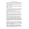
A new page displays your results. Scroll down to the Alignments section to see the two sequences aligned. The reference sequence will be on top (Query), and the patient 1 sequence will be below it (Sbjct). The screen should look similar to Figure 1 below.
Note that identical nucleotides are indicated with a vertical line drawn between them. Scan the alignment to find any nucleotides that are not identical, and write down the nucleotide number and the nucleotides in the reference and patient sequences at that number.
Note: The only way to determine the numerical location of a SNP is to count from one end of the line or the other. The number of the first nucleotide on each line is shown to the left of the line, and the last number is shown to the right of the line. In the standard BLAST output, each line contains 60 nucleotides. To determine the precise location of a SNP, you must count carefully from one end of the line or the other. In Figure 1, the location of the SNP is at nucleotide 24, which has an "A" in the patient sequence (Sbjct) and a "G" in the reference sequence (Query).
Return to the BLAST page by clicking the Edit and Resubmit link in the upper left. Clear the Enter Subject Sequence box by clicking “Clear” above the box. Then repeat steps 4-8 for the patient 2 sequence. (You may leave the Enter Query Sequence box unchanged, as it will still contain the reference sequence.)
Figure 1. Example BLAST results
BLAST Search Instructions
In the middle of the page, under the Basic BLAST heading, click nucleotide blast. A new page will appear.
In the Enter Query Sequence panel, check the box next to Align two or more sequences, and wait for the page to refresh.
Copy the reference sequence shown above, and paste it into the Enter Query Sequence box at the top of the page.
Copy the sequence for patient 1, and paste it into the Enter Subject Sequence box.
In the Program Selection panel, select Highly similar sequences (megablast).
Click the BLAST button near the lower-left corner of the page. Wait for BLAST to complete the alignment.
A new page displays your results. Scroll down to the Alignments section to see the two sequences aligned. The reference sequence will be on top (Query), and the patient 1 sequence will be below it (Sbjct). The screen should look similar to Figure 1 below.
Note that identical nucleotides are indicated with a vertical line drawn between them. Scan the alignment to find any nucleotides that are not identical, and write down the nucleotide number and the nucleotides in the reference and patient sequences at that number.
Note: The only way to determine the numerical location of a SNP is to count from one end of the line or the other. The number of the first nucleotide on each line is shown to the left of the line, and the last number is shown to the right of the line. In the standard BLAST output, each line contains 60 nucleotides. To determine the precise location of a SNP, you must count carefully from one end of the line or the other. In Figure 1, the location of the SNP is at nucleotide 24, which has an "A" in the patient sequence (Sbjct) and a "G" in the reference sequence (Query).
Return to the BLAST page by clicking the Edit and Resubmit link in the upper left. Clear the Enter Subject Sequence box by clicking “Clear” above the box. Then repeat steps 4-8 for the patient 2 sequence. (You may leave the Enter Query Sequence box unchanged, as it will still contain the reference sequence.)
Figure 1. Example BLAST results
Go to Question 1
Section II. How does each SNP affect the amino acid sequence of the insulin protein?
In Part I you identified the location and base substitution of all four patients’ SNP. The next step is to determine how these base substitutions alter the amino acid sequence of the insulin protein. The wild-type nucleotide sequence of the insulin cDNA is shown in Figure 2 (top line). Below each triplet of nucleotides is the amino acid specified by that codon. (Nucleotide numbers are blue, and amino acid numbers are red.)
Recall that the flow of genetic information is as follows: DNA RNA Proteins. The cDNA nucleotide sequence shown in Figure 2 corresponds to the coding (nontemplate) strand, so to convert it to mRNA for use with the codon table, you only need to change T to U.
Figure 2. Wild-type insulin cDNA sequence and corresponding amino acids
For each patient, use the information in Figure 2 to identify the wild-type amino acid at each SNP location, then use the codon table (page 127 of your book) to determine the new amino acid at the SNP location. Finally, determine the type of mutation (e.g., silent, missense, nonsense) caused by each SNP. Patient 1 has been completed for you, as shown in Question 2 of your assignment.
HINTS
Hint 1. What do the numbers and letters in the “SNP (DNA)” column mean?
The combination of numbers and letters in the SNP (DNA) column are a standard shortcut for describing any SNP. It is written in this sequence:
the nucleotide in the wild-type sequence (A, T, G, C)
the numerical position of the SNP in the sequence (number)
the nucleotide that is present in the SNP (A, T, G, C)
For example, the designation T125G means that a thymine (T) in the wild-type sequence at position 125 has been replaced by a guanine (G) in the SNP.
Hint 2. How do you figure out which amino acid is encoded by the SNP?
Use the following steps to fill in the table in Question 2. We’ll use Patient 1 as an example:
Find the location of the SNP in the wild-type insulin cDNA sequence. In Patient 1, this is nucleotide number 125, which is a T in the wild-type sequence.
Identify the wild-type triplet codon that the SNP is located in, and the corresponding amino acid. In this example, the codon is GTG, which encodes a Val (valine) amino acid.
Note that cDNA information in the table corresponds to the cDNA coding (nontemplate) strand, so to convert it to mRNA for use with the codon table, you just need to change T to U.
Determine the new codon in the SNP and the corresponding amino acid. In this example, the T in the wild-type sequence has been replaced by a G in the SNP. Thus the new codon is GGG. According to the codon table, GGG encodes a Gly (glycine) amino acid.
Hint 3. What distinguishes different types of mutations?
The type of mutation that results from a SNP depends on the difference between the amino acid encoded by the wild-type codon and the amino acid encoded by the new codon produced by the SNP. The point mutations are described on page 135 of your book.
Go to Questions 2 and 3
Section III. Which domain of the insulin protein is affected by each SNP? What are its effects?
In Part II you identified the amino acid change in the insulin protein caused by each SNP. However, not all amino acids in the translated insulin protein contribute equally to the function of the final insulin protein. Next you will learn about processing of the insulin protein and how SNPs located in different regions of the protein can have profoundly different effects on insulin function.
Translation of the insulin mRNA produces a precursor protein of 110 amino acids, shown in Figure 3 below. There are five different regions (or “domains”) of the insulin precursor protein, including the (1) signal sequence, (2) dibasic site, (3) C peptide, (4) A chain, and (5) B chain.
A mature insulin protein is created with the following details in mind…A signal sequence (brown) at the N-terminal end of the insulin precursor protein targets the protein to the ER. In the ER, the signal sequence is cleaved off, and three disulfide bonds are formed between pairs of Cys residues. Proteases (enzymes that break down proteins and peptides) then cleave the precursor protein at two sites, each adjacent to a pair of basic amino acids (called dibasic sites, green), releasing the C peptide (blue). Finally, the dibasic sites are removed. This generates the mature insulin protein, consisting of the A chain (yellow) and B chain (purple) joined by a pair of disulfide bonds. It is this mature form of the insulin protein that is secreted into the blood.
Precursor protein with 110 amino acids
Mature insulin protein with 51 amino acids
Precursor protein with 110 amino acids
Mature insulin protein with 51 amino acids
Figure 3. The structure of the precursor insulin protein and the mature insulin protein
It is reasonable to assume that any changes in the normal domain structure of wild-type insulin are likely to disrupt function. However, it should be noted that SNPs can have profoundly different effects on the function of the mature insulin protein depending on which domain they are found in. For example, SNPs in the dibasic site domain block the removal of the C peptide and render the protein nonfunctional. Similarly, SNPs that alter the signal sequence would prevent subsequent processing and render the protein nonfunctional. Contrastingly, SNPs that alter amino acids in the C peptide are unlikely to affect insulin structure or function (unless they somehow prevent processing) since the C peptide domain is cleaved off and not included in the mature insulin protein. Finally, the effect of SNPs in the A and B chains is variable. This is because we cannot predict if the substitutions (taken collectively) will cause large changes in structure or not. The exception is substitutions for the four Cys residues that are involved in disulfide bond formation. Preventing the formation of either disulfide bond radically affects structure and thus function. Further tests would be needed to confirm the effect of SNPs in the A and B chains that do not affect the Cys residues.
Use this information to draw conclusions about the functioning of the mature insulin protein in patients 1 through 4. Note that patients who have a “nonfunctioning protein” as described above, exhibit diabetes.
Go to Questions 4 and 5
Part B. Investigating Genetic Disorders
In this section you’re tasked with researching a genetic disorder of interest to you (or assigned to you upon request). There are a number of websites and textbooks you can visit to obtain information on disorders. I’ve provided three websites below to start your investigation – please keep a list of the informational sources you use, and list them at the end of this assignment.
Respond to questions 1 through 5 in the Part B section of your assignment questions. Note: in answering the questions, you must use your OWN words. You may be asked to share your research with your classmates and I expect you will be able to “teach” your peers about the disorder that you researched.
Resources:
University of Utah Health Sciences: http://learn.genetics.utah.edu/content/disorders/
Cold Spring Harbor Laboratory: http://www.ygyh.org/
U.S. Department of Energy Human Genome Project: http://web.ornl.gov/sci/techresources/Human_Genome/posters/chromosome/
Assignment 4 Questions
Part A. Which Insulin Mutations May Result in Disease? [20 pts]
Using the results of your BLAST alignments, complete the table below. 3 pts
SNP location (nucleotide no.)
Wild-type nucleotide at this location
Nucleotide present in patient as a SNP
Example (Fig 1)
24
G
A
Patient 1
125
T
G
Patient 2
220
G
A
Patient 3
164
G
C
Patient 4
309
C
G
For each patient, use the information from question 1 to determine the SNP (DNA) (see Hints in the instructions). Then use Figure 2 to identify the wild-type amino acid at each SNP location, and the codon table (page 127 of your book) to determine the new amino acid at the SNP location. Finally, determine the type of mutation (e.g., silent, missense, nonsense) caused by each SNP. Patient 1 has been completed for you. 6 pts
SNP (DNA)1
Wild-type amino acid at SNP location
New amino acid at SNP location
Mutation type
Patient 1
T125G
Val
Gly
Missense
Patient 2
G220A
Ala
Thr
Missense
Patient 3
G164C
Arg
Pro
Missense
Patient 4
C309G
Tyr
STOP
Nonssense
1 Review the “Hints” box for information about what the numbers and letters in this column mean.
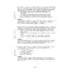
Describe how protein structure and function will differ as a result of a silent vs. missense vs. nonsense mutation. 3 pts
In silent, the amino acid doesn’t change, therefore the protein structure doesn’t change. In missense, the amino acid changes from one to another therefore the proteins will differ often becoming mutant. In nonsense, the amino acid changes into a stop codone which in turn changes the protein into a somewhat defective and short one.
Which domain of the insulin precursor protein is affected by each of the four SNPs? Hint: the “amino acid position” can be found by locating the SNP (DNA) and the corresponding amino acid (location) associated with the SNP (see Figure 2). The domain in which each amino acid is located can be found by examining Figure 3. 4.5 pts
Amino acid position
Domain affected by SNP
Expected impact of SNP on the insulin protein formation
Patient 1
42
B chain
Variable effect on insulin
Patient 2
75
C peptide
Unlikely to affect insulin
Patient 3
55
Dibasic Site
Nonfunctional protein hinders insulin
Patient 4
103
A chain
Variable effect on insulin
Of the four patients whose SNPs you investigated, which are at risk of diabetes and which ones are not? Explain your response. 2 pts
Patient 3 is at risk of diabetes because thw SNP was at the dibasic site. When changed, the dibasic site cannot block the removal of the C chain and peptide.
What did you learn from this exercise about genetic mutations and the formation of proteins? 1.5 pts
I learned that having some type of genetic disorder is much more common than I realized because all that has to happen is for one set of proteins to not match up or to be missing.
Part B. Investigating Genetic Disorders [10 pts]
What genetic disease did you select? 0 pts
Down syndrome
What causes the disease? Here please focus on the genetic basis of this disease. Questions to address (though not exclusively): Is the disease caused by a dominant or recessive gene allele? What chromosome is the gene found on? Is the disease caused by defects in more than one gene? Does the environment play a role? Genes contain the information to make proteins, so you will likely find information both on the defective gene and what is known about the defective protein. What is the protein supposed to do? What does it do in a person with the disease? 7 pts
Down syndrome is a disorder caused by cell division issues. There ends up being an extra chromosome.
Down syndrome is NOT passed down through alleles. The mutation is more likely to occur when mothers over 35 are having children.
The sperm and egg come together and the resulting embryo has a third copy of chromosome 21.
No gene on the chromosome is abnormal, the only difference is that there are three of #21.
There are no known environmental factors that cause down syndrome.
Chromosome 21 has 200-300 genes that have instructions to make proteins for the rest of the body. This causes proteins without the body to either be under or overexpressed.
How common is the disease? Are certain people more likely to get it than others, and why? 1 pt
As a very common birth defect, about 6000 babies born each year in the US have down syndrome (March of Dimes). If a mother is older than 35 when pregnant, her child is more likely to have down syndrome.
What are its symptoms? When does it strike? 1 pt
Down syndrome can be noted with particular facial features (flat face, upward-slanted eyes, small, broad nose, oddly shaped ears). There can also be the aspect of a moderate to severe intellectual disability. Development of walking, talking, and taking care of themselves is slower in comparison to others. Doctors can screen for down syndrome by taking a sample from the placenta, amniotic fluid, or umbilical cord to examine the baby’s chromosomes.
What cures or treatments are available or in development for this disease? 1 pt
There is no cure although different therapies such as physical and speech can help with developmental problems.