Transcript
Chapter 5: Membranes
Introduction
Membrane components
Lipids
Proteins
Carbohydrates
Membrane function
Types of transport across membranes
Diffusion
Facilitated diffusion
Active transport
Exo- and endocytosis
1. Membranes are a mosaic of lipids, proteins and carbohydrates (Fig. 5.1)
Membranes are fluid: the components move.
Membranes are spatially variable.
Cells regulate and change the components of their membranes over time.
2. Membrane components
2a. Phospholipids are the most common lipid and the bulk of the membrane (Fig. 3.12)
In an aqueous solution, phospholipids spontaneously form bilayers (5-10 nm thick) and micelles (Fig. 2.17).
There are different types of phospholipids.
Head groups differ in size, shape, charge
Fatty acid length and saturation differs among cell types and different membrane areas
Changing lipids changes membrane characteristics.
Shorter hydrocarbon tails = thinner membrane, greater membrane fluidity.
More unsaturated fatty acids = greater membrane fluidity
Phospholipids drift laterally; need energy and a protein to flip to other layer (Fig. 5.3).
Animal cells have cholesterol in their membranes.
Cholesterol stabilizes membranes.
Makes it less fluid at high temperatures, more fluid at low temperatures.
2b. Proteins give the membrane function.
How proteins associate with membranes:
Integral membrane proteins:
1. Transmembrane proteins: physically inserted into the membrane.
2. Lipid-anchored proteins: amino acid and R group is covalently bound to a membrane lipid.
Peripheral membrane proteins:
Noncovalently bound to other membrane proteins or to a phospholipid polar head.
Proteins are big, so move slowly through membranes. If they are linked to other molecules, they may not move at all (Fig. 5.5)
Cells regulate and change the protein content of their membranes.
About 25% of all genes encode for membrane proteins
2c. Carbohydrates may be covalently attached to proteins or lipids (= glycosylation).
Roles of glycosylation:
Cell: cell recognition
Protection of cell membrane from physical damage and proteases (an enzyme that digests proteins).
Carbs coat a white blood cell (Fig. 5.6)
Glycoproteins and glycolipids only on the external surface membrane bilayers (= leaflets) have different components.
3. Membrane function
It is a barrier between environment and cell.
Selectively permeable, depending on molecule’s
Size
Charge
Transmembrane concentration gradient
Hydrophilic nature
4. Types of transport across membranes
4a. Diffusion: substance moves from a region of high concentration to a region of low concentration. A type of passive transport.
Small, nonpolar molecules easily diffuse (or, the membrane is highly permeable).
Ions and large molecules: very low permeability.
Ion transport across membranes can drive
ATP production
Transmembrane movement of some solutes
Production/transmission of electrical signals
Osmosis: diffusion of water across a semipermeable membrane to balance solute concentrations.
Tonicity: the relative solute concentration
1. Hypertonic environment: more concentrated than the cell.
2. Isotonic environment: equal concentration to the cell.
3. Hypotonic environment: less concentration than the cell.
Can also describe cell relative to environment.
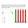
The effect of varying tonicity (Fig. 5.16a)
Hypertonic environment: cells shrink
Isotonic environment: no volume change
Hypotonic environment: cells swell
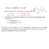
4a. Passive diffusion (no transport protein)- movement of high concentration to low concentration
Fick equation (p.839) determines rate of diffusion across a membrane. J= KA(C1-C2)
J= rate of diffusion
K= constant (includes temp.)
A= cross-sectional membrane area
C= concentration at a location
4b. Facilitated diffusion: a protein that increases selective permeability (Fig. 5.11b)
#2a: a channel: an open passageway that spans the membrane and doesn’t use energy.
Example: Aquaporin protein channels facilitate diffusion of water (Fig. 5.20)
Channels may be gated: open and close in response to a signal. (Fig. 5.21)
Signals may be chemical or physical.
Transporters are proteins that facilitate diffusion by binding solutes and changing shape (Fig. 5.22).
4c. Active Transport: use ATP and protein transports to move solutes against gradient.
4d. Large particles are taken in by endocytosis and released by exocytosis.