The
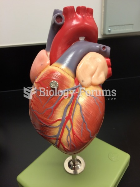
pacemaker cells of the heart are involved in originating and conducting action potentials through the heart. The conduction pathway starts at the sinoatrial node, where most cardiac cycles are initiated. The heart is said to have an autorhythmicity because it is able to generate its own action potential. The pacemaker cells of the heart are responsible for this autorhythmicity, because they spontaneously generate their own action potentials. The source of that capacity resides with the ion channels that are active within pacemaker cells. After the resting membrane potential is reached, the K+ channels that were activated to return membrane potential to control will close, such that the membrane begins to slowly depolarize. At the same time, a channel that is relatively non-specific for K+ or Na+ (a funny channel, non-selective cation channel) begins to open, furthering the slow depolarization of the membrane. Before membrane potential can reach threshold, the T-type Ca2+ channels are activated. They will further the depolarization. These channels are only open for a short time span, but the depolarization that they mediate stimulates the opening of L-type Ca2+ channels that are responsible for the action potential. In the pacemaker cells, the action potential is driven by the influx of Ca2+ rather than Na+. The stimulation of L-type Ca2+ channels is regenerative, as is the stimulation of Na+ channels in neurons and skeletal muscles. The opening of one channel will induce a depolarization that stimulates others to open, further depolarizing the cell's membrane. At some point, the permeability of that membrane to K+ is elevated. That elevation in K+ permeability is voltage dependent but occurs more slowly than the opening of L-type Ca2+ channels. The frequency with which pacemaker cells develop an action potential is affected by the rate of depolarization. The faster the cells depolarize, the sooner the cell will reach threshold and an action potential will be generated.
 Quick Reply