Transcript
Biology 20
Muscular System Notes
Chapter 10
Name:____________________
Muscles and More
Goals for this Chapter:
Observe and compare the three types of muscle tissue
Describe the action of actin and myosin in muscle contraction and heat production
Identify the sources of energy for muscle contraction
Explain how skeletal muscles support other body systems
Identify conditions that impair the healthy functioning of muscle systems
Describe the benefits of exercise for maintaining healthy muscles
Text book Pg 330-359
Magnificent Muscle Facts
The human body has more than 650 muscles
Waste energy keeps you warm!
No two muscles in the body have exactly the same function. When one muscle is paralyzed, either stability of the body part is impaired or some specific movement is lost.
A muscle is pretty efficient, using about 35-50% of its potential energy
Muscle fibers are thinner than a human hair and can support up to 1,000 times its own weight.
The strongest muscle in the human body is masseter?
Muscle is Latin for “little mouse”
MY Oh MY Muscles
Types of Muscles
286385-381000
43688003365500
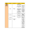
Types of muscle
Skeletal muscle
Striated & tubular
Many nuclei
Voluntary
Attached to bones
Functions in circulatory system to help move blood back to the heart (contract against veins)
Release heat
Move bones
Allow us to stand upright
350964517716500Cardiac muscle
Striated (little lines in muscle), tubular and branched.
One nucleus
Involuntary control
Found only in walls of heart
______________________
Smooth muscle
45161201968500Non-striated
One nucleus
Involuntary control
Found in walls of internal organs
Ex: sphincter muscles of esophagus, intestines, and urethra
Does not fatigue easily
Muscular System Supports the Skeletal System
1631953429000
Action of Muscles
During contraction a muscle shortens when stimulated by an excitatory nerve
During relaxation a muscle lengthens when stimulated by an inhibitory nerve
Muscles Cooperate
Muscles can only pull therefore they must work in pairs pulling against each other (antagonistic)
Eg. Biceps-triceps
Bicep pulls to bend a joint (___________)
Tricep pulls to straighten a joint (___________)
308610011430000-11430011430000
Muscle Type
Location in Body
Structure
Function
Cardiac Muscle
Smooth Muscle
Skeletal Muscle
Muscle Organization
Muscle
An organ surrounded by connective tissue and composed of several tissues
eg. bundles of muscle cells and nervous tissue.
Muscle fibres
muscle cells in the bundles
unlike “typical”cells.
Up to 20 cm in length
contain many nuclei
Organelles within the cells (fibres)
3171520118515the cell membrane is called the sarcolemma
333028055580the endoplasmic reticulum is the sarcoplasmic reticulum
5987850-293155886330149655832330-12795557210905312556163304052554798906572552066501103654755930786854937010-87547717705636545939301625654225650234205413673018020539560101611253784290199285385737014204534955702244852968530310165278128065725the cytoplasm is known as sarcoplasm.
2508150-1206902295390-479701752510-922501390350-47970790230-85770631470-76410498270-54090367590-25650266430-38250110910-44730283464012382500012382500
Skeletal Muscle Structure
Muscle fibres (a specialized cell) are organized into larger bundles up to 20 cm in length
Connective tissue wraps fibref
Another layer of connective tissue bundles fibres
Muscle Structure
Myofibrils are composed of small subunits called sarcomeres
Sarcomeres are the functional unit of a muscle between z disc (zigzaggy)
Composed of actin and myosin (protein chains)
Actin is the thin filament
Myosin is the thick filament
3263906413500
40322521018500
F
Skeletal Muscle Structure
Blood vessels and nerves run between bundles or fibres
Cell membranes of muscle cells are called sarcolemma
Fibres are composed of myofilaments
Myofilaments can be thin (actin filament) or thick (myosin filament) and overlap to give alternating light and dark bands.
Muscle Contraction
One sarcomere is composed of both actin (thin) and myosin (thick) filaments
_____________ divide sarcomeres
_____________ is the area where myosin can be seen
Dark areas are A-bands (myosin & actin)
Lighter areas are I-bands (actin)
Muscle contraction can be explained by the Sliding Filament Theory
The lengths of thick filaments (myosin) and thin filaments (actin) do not change during contraction.
The current hypothesis is that the thin and thick filaments slide past each other during contraction
Sliding Filament Theory
Sliding Filament Theory
Signal to muscle from a nerve
Sarcoplasmic reticulum releases stored Ca2+
Ca2+ binds to troponin which in turn moves tropomyosin (protein that covers the binding sites for myosin heads) allowing the myosin to bind
calcium is returned to the sarcoplasmic reticulum through active transport when the contraction stops
Links:
http://www.youtube.com/watch?v=gJ309LfHQ3M
http://www.youtube.com/watch?v=0kFmbrRJq4w
Energy for Muscles
There are three main sources for muscles:
Stored creatine phosphate
Anaerobic respiration
Aerobic respiration of glucose and fatty acids
Muscle fibre contraction is paid for using ATP
After a few muscle twitches ATP is spent in the muscles.
The ATP that is used afterwards comes from the 3 sources listed above.
6686559271000
High energy molecule
found in amongst the sliding filament
adds Pi to ADP to replenish the ATP being consumed in the muscles
consumed in 8 secs
replenished during rest
Slow Twitch Muscles – Type I
Takes longer to contract (100 ms for tetanus)
Contains myoglobin (stores oxygen in muscle) gives red colour to muscle, more blood, more mitochondria
ATP produced aerobically
(sports like long distance running, swimming, biking)
in size with training
Fast Twitch Muscles – Type II
Fast contractions (7 ms for tetanus)
Anaerobic ATP generation
Glycogen rich, light colour
Fatigue quickly (vulnerable to lactate)
Sports like sprinting, weight lifting
Eye movements use fast twitch contractions
Increase in thickness with training
Intermediate – Type IIa
Fast twitch
Increased aerobic capacity
Formed from endurance training
10.2 – Muscles, Health, and Homeostasis
Even at rest, muscles are still contracting at some level
We rely on proper muscle tone to maintain our posture, and to keep us upright
Complications of the Muscular System
Muscles are generally vulnerable to injuries that result from sudden stress
However, muscles are one of the few organ groups whose activity can be impaired through lack of use
Muscular atrophy results from a lack of movement of the muscle
Common Muscular Disorders
Muscular Dystrophy-
Botulism-
Fibromyalgia-
Cramps
Contracture
Crush syndrome
Delayed onset muscle soreness
Myositis
Common Injuries to Muscular System
Torn muscles
Stretched tendons
Torn ligaments
Joint sprains
Joint dislocation
Exercise & Muscle Contraction
Regular exercise allows muscles to develop and use energy more efficiently
Regularly used muscles grow due to the increase in the size of the individual muscle fibre, not because of the increase in the number of fibres
The increase in the size of muscles is known as hypertrophy
Homeostasis
Our muscular system allows us to maintain homeostasis
Our muscles generate heat through the use of ATP during contraction, and muscles allow blood vessels to contract and dilate to move warm blood throughout the body
As well, many of our processes in our other body systems rely on the movement of muscles to regulate actions
Relationship to Other Systems
Relationship
Circulatory System
Respiratory System
Digestive
System
Excretory
System
1. Three types of muscle tissue are located in various organs in the body. Complete the table below to summarize the characteristics of each type of muscle tissue.
Characteristics of Muscle Tissues
Type
Structure
Voluntary/ Involuntary?
Location in the Body
Smooth
Cardiac
Skeletal
Use the following information to answer the next question.
Although they differ in various characteristics, all muscles types have one function. They create movement within the body.
Review the figure opposite. It explains that sensors in the body continuously detect stimuli. Information is sent to control centres, which in turn direct responses to the stimuli. Responses are carried out by effectors—all of which are types of muscle tissue.
Suppose you are asleep and the doorbell rings. You stumble to your feet and move toward the door, open it and speak with your visitor. Think of the many changes occurring in your body as you awaken and respond to the sound of the doorbell.
2. Complete the table below to show how the three types of muscle tissue help you respond to the doorbell.
Smooth Muscle
Cardiac Muscle
Skeletal Muscle
Use the following information to answer the next two questions.
Normally, skeletal muscle contractions are stimulated and controlled by nerve cells. Calcium ions (Ca2+) also play an important role in the way muscles are controlled, as demonstrated by a series of experiments using isolated muscle fibres. These fibres can be manipulated in various ways:
They can be stimulated with electrodes to mimic the effect of nerve cells.
Ca2+ in solution can be injected into the fibres.
A chemical that removes Ca2+ already present in the fibres can be injected.
The results from experiments using these procedures are given in the table below.
The Effect of Calcium Ions on Muscle Fibre Contraction
Experiment
Procedures
Results
Electrode Stimulation
Ca2+ injected or removed
1
on
neither
contraction
2
off
injected
contraction
3
on
removed
no contraction
1. Interpret the results of these experiments.
2. What evidence is there from these experiments that calcium ions are normally present in muscle tissue?
Use the following additional information to answer the next four questions.
Additional experiments using injections of radioactive Ca2+ show that the ions are stored within the sacs of the sarcoplasmic reticulum in resting muscle tissue. When the tissue is stimulated to contract with electrodes, the radioactive Ca2+ ions are found among the actin and myosin filaments as shown below.
3. Refer to diagram of the muscle at rest above, and explain what effect a lack of tropomyosin would have in muscle tissue.
4. The diagram of the muscle contracting shows the role of calcium ions in repositioning tropomyosin. Where are these ions stored when the muscle is at rest? What causes them to move among the actin and myosin filaments?
5. What happens to calcium ions and tropomyosin to cause a muscle to relax?
6. Use a flow chart to describe the series of events that starts with stimulation and ends with relaxation.
Once energy from ATP and creatine phosphate stored in working muscle fibers is exhausted, ATP is generated from the breakdown of glucose and fatty acids through aerobic and anaerobic respiratory pathways. To keep contracting, the muscle increases aerobic cellular respiration and carries out fermentation as oxygen becomes scarce.
In the contracting muscle, ATP is broken down to ADP + P as energy is spent on movement and heat is released.
1. Explain how red muscle is well adapted to accommodate a high rate of cellular respiration.
2. Involuntary shivering occurs when body temperature decreases. Why is this response an advantage for survival?
3. Recalling your knowledge of the circulatory system, explain how wasted heat from strenuous exercise is dissipated from the body.
4. In fermentation, ATP is generated without oxygen. It may seem that this involves getting something for nothing. Is this true? Explain.