|
Description
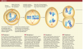
The stages of meiosis. In this form of cell division, replicated homologous chromosomes physically associate to form a chromosome pair. Members of each chromosome pair separate from each other at the first meiotic division (meiosis I). In meiosis II, the centromeres of unpaired chromosomes divide, resulting in four cells, each with the haploid (n) number of chromosomes.
(a) Prophase I
At the end of interphase, chromosomes are duplicated and in threadlike form. Now they start to condense. Each pairs with its homologue, and the two usually swap segments. The swapping, called crossing over, is indicated by the break in color on the pair of larger chromosomes. Newly forming spindle microtubules become attached to each chromosome.
(b) Metaphase I
Motor proteins projecting from the microtubules move the chromosomes and spindle poles apart. Chromosomes are tugged into position midway between the spindle poles. The spindle becomes fully formed by the dynamic interactions among motor proteins, microtubules, and chromosomes.
(c) Anaphase I
Some microtubules extend from the spindle poles and overlap at the equator. These lengthen and push the poles apart. Other microtubules extending from the poles shorten and pull each chromosome away from its homologous partner. These motions move the homologous partners to opposite poles.
(d) Telophase I
Cytokinesis divides the cytoplasm of the cell after telophase. There are now two haploid (n) cells with one of each type of chromosome that was present in the parent (2n) cell. All chromosomes are still in the duplicated state.
|